
AP Biology students will relearn mitosis and cell division, though with older students, I like to add more challenging concepts. In this exercise, students read about research conducted by Leonard Hayflick, which lead to the discovery that cells have a lifespan based on the number of cell divisions.
The student worksheet explores the discovery of this phenomenon and the role of telomeres in cell division and aging.
The Hayflick limit, refers to the number of times a normal somatic cell can divide before cell division stops. Cell division and senescence: Hayflick demonstrated that normal human fetal cells can divide between 40 and 60 times in cell culture before entering a phase called senescence
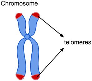
The Hayflick limit is also closely related to the shortening of telomeres, which are protective caps at the ends of chromosome Each time a cell undergoes mitosis (cell division), the telomeres shorten. Once the telomeres reach a critical length, cell division cannot occur.
Telomere shorting is the likely cause of cell senescence. With each cell division, telomeres become shorter. Eventually the cell no longer can divide and will die (apoptosis). This type of programmed cell death removes older cells that may have accumulated mutations.
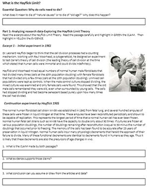
After students read the experiments, they highlight statements that indicate CLAIMS made by the researchers, and then highlight evidence statements. My students are familiar with the CER format.
In the final section, students analyze a graph that shows cell counts from normal gastric cells. These are compared to cell counts from cancerous cells. The graph indicates that cancerous cells will continue to divide.
Students are also introduce to the concepts of telomerase, which is an enzyme that can reverse the shortening of telomeres and increase the lifespan of cells. Telomerase is one way that cancer cells can bypass the Hayflick limit and become “immortal.”
I usually do this activity after students have completed the mitosis investigation. In this activity, students look at dividing cells using a microscope and estimate the time spent in each stage of the cell cycle.