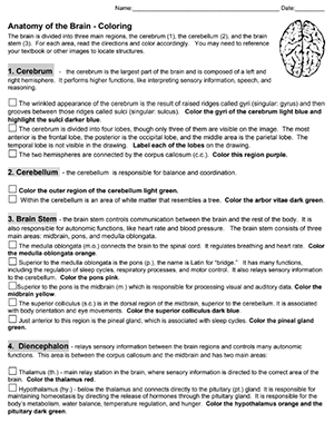
Students learn brain structures and their functions by reading short descriptions and coloring an image.
The description are organized into three main areas of the brain: cerebrum, cerebellum, and brain stem. The brain stem also includes structures of the diencephalon (thalamus and hypothalamus).
In addition to coloring the image according to the directions, students identify which parts of the brain are related to a list of functions. The answers can be found within the text of the document. Students may also refer to their notes on the brain to help with this exercise.
I have quite a few brain labeling activities that I use with my anatomy class, as well as a sheep brain dissection. This coloring exercise was made for students who need additional reinforcement or who could not complete the dissection.