Explore the Anatomy of the Brain with Coloring
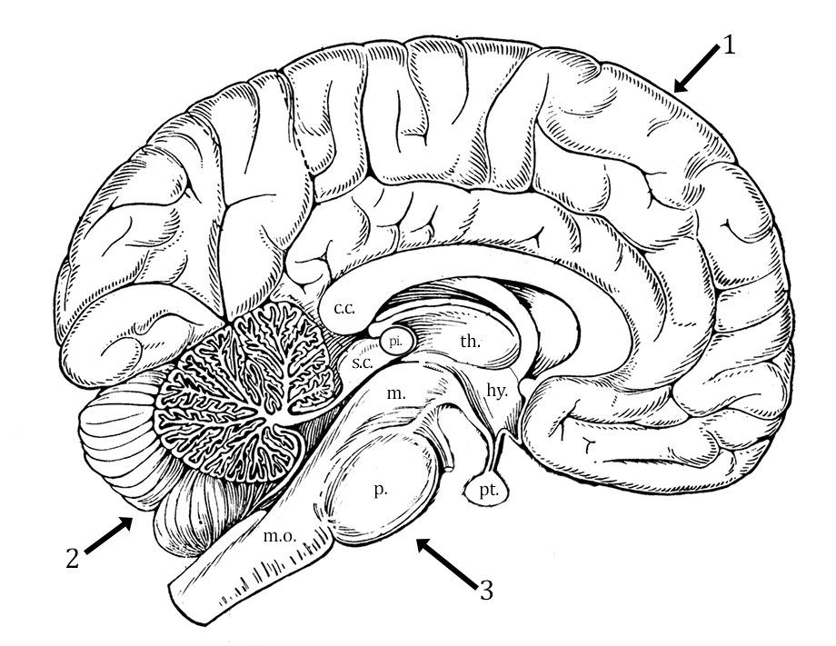
 The brain is divided into three main regions, the cerebrum (1), the cerebellum (2), and the brain stem (3). For each area, read the directions and color accordingly. You may need to reference your textbook or other images to locate structures.
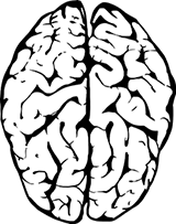
1. Cerebrum - the cerebrum is the largest part of the brain and is composed of a left and right hemisphere. It performs higher functions, like interpreting sensory information, speech, and reasoning.
 The wrinkled appearance of the cerebrum is the result of raised ridges called gyri (singular: gyrus) and then grooves between those ridges called sulci (singular: sulcus). Color the gyri of the cerebrum light blue and highlight the sulci darker blue.
The cerebrum is divided into four lobes, though only three of them are visible on the image. The most anterior is the frontal lobe, the posterior is the occipital lobe, and the middle area is the parietal lobe. The temporal lobe is not visible in the drawing. Label each of the lobes on the drawing.
The two hemispheres are connected by the corpus callosum (c.c.). Color this region purple.
2. Cerebellum - the cerebellum is responsible for balance and coordination.
Color the outer region of the cerebellum light green.
Within the cerebellum is an area of white matter that resembles a tree. Color the arbor vitae dark green .
3. Brain Stem - the brain stem controls communication between the brain and the rest of the body. It is also responsible for autonomic functions, like heart rate and blood pressure. The brain stem consists of three main areas: midbrain, pons, and medulla oblongata.
The medulla oblongata (m.o.) connects the brain to the spinal cord. It regulates breathing and heart rate. Color the medulla oblongata orange.
Superior to the medulla oblongata is the pons (p.), the name is Latin for “bridge.” It has many functions, including the regulation of sleep cycles, respiratory processes, and motor control. It also relays sensory information to the cerebellum. Color the pons pink.
The superior colliculus (s.c.) is in the dorsal region of the midbrain, superior to the cerebellum. It is associated with body orientation and eye movements. Color the superior colliculus dark blue.
Just anterior to this region is the pineal gland, which is associated with sleep cycles. Color the pineal gland green.
4. Diencephalon - relays sensory information between the brain regions and controls many autonomic functions. This area is between the corpus callosum and the midbrain and has two main areas:
Thalamus (th.) - main relay station in the brain, where sensory information is directed to the correct area of the brain. Color the thalamus red.
Hypothalamus (hy.) - below the thalamus and connects directly to the pituitary (pt.) gland. It is responsible for maintaining homeostasis by directing the release of hormones through the pituitary gland. It is responsible for the body’s metabolism, water balance, temperature regulation, and hunger. Color the hypothalamus orange and the pituitary dark green.
Identify the part of the brain responsible for:
1. Sleep cycles (gland) __________________________________________
2. Reasoning __________________________________________________
3. Balance and coordination _______________________________________
4. Connecting left and right hemisphere ____________________________________
5. Visual and auditory data processing ______________________________________
6. Regulation of breathing and heart rate ____________________________________
7. Maintaining homeostasis by directing the pituitary ___________________________
8. Body orientation and eye movements ____________________________________
9. Relays information to the correct area of the brain ______________________________
10. Regulates sleep cycles, respiratory processes and motor control ___________________________