
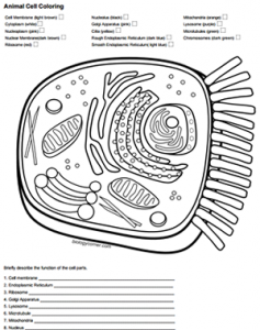
This worksheet requires students to color a drawing of animal cell according to directions. Students may need to use their book or other resources to identify parts of the cell, like the mitochondria, golgi apparatus, endoplasmic reticulum, and ribosomes.
This activity is a simple reinforcement worksheet to help students learn the structures found within a typical animal cell and what those structure might look like on a diagram. You can also assign this with the plant cell coloring.
Student can practice labeling skills with Simple vs. Complex Cell diagrams.
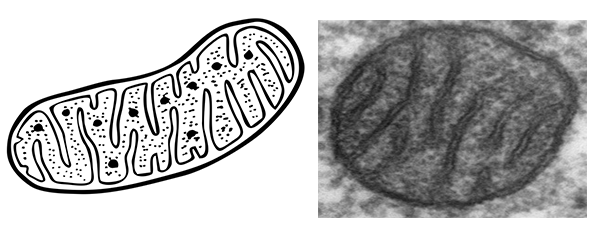
Students are sometimes confused by diagrams that don’t resemble the ones they have seen in their textbook. I stress to my students that these drawings are just representations of what an artist imagines the organelle looks like.
Most organelles will look similar in the diagrams, but they are all interpretations of microscopic views. Most of our understanding about what the mitochondrion looks like is from transmission electron microscopes.
HS-LS1-2 Develop and use a model to illustrate the hierarchical organization of interacting systems that provide specific functions within multicellular organisms