
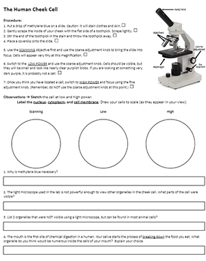
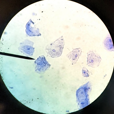
In this lab, students use a toothpick to get a sample of cells from the insides of their cheek. These cells are stained with methylene blue and observed with a light microscope. Cells can be seen clearly at low power (100x) though they will be very small. Focusing on high power will show more details of the cell, though generally the only structure large enough to be visible is the nucleus.
Students can also view skin cells by applying a piece of clear tape to their skin and then placing the tap on the microscope (no cover slip needed).
Worksheet contains simple instructions for focusing the microscope, though this lab is usually completed after students have done the introductory microscope lab or with students in second year biology or anatomy.
In my anatomy class, I also provide prepared slides of tissues for students to view when they are finished with the cheek cell. These include slides showing skin, blood, and even muscle.
Cheek cells can be challenging, encourage students to move the slide around to find a good sample spot. Air bubbles are often mistaken for cells. My students share their cheek cells by snapping a picture with their cell phone and posting to twitter, #cellfie.
Grade Level: 7-12 | Time Required: 30 – 45 min
HS-LS1-2 Develop and use a model to illustrate the hierarchical organization of interacting systems that provide specific functions within multicellular organisms