The Human Cheek Cell


Procedure:
1. Put a drop of methylene blue on a slide. Caution: methylene blue will stain clothes and skin.
2. Gently scrape the inside of your cheek with the flat side of a toothpick. Scrape lightly.
3. Stir the end of the toothpick in the stain and throw the toothpick away.
4. Place a coverslip onto the slide.
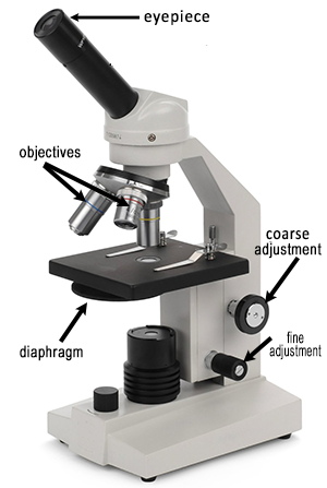
5. Use the SCANNING objective first and use the coarse adjustment knob to bring the slide into focus. Cells will appear very tiny at this magnification.
6. Switch to LOW POWER and use the coarse adjustment knob. Cells should be visible, but they will be small and look like nearly clear purplish blobs. If you are looking at something very dark purple, it is probably not a cell.
7. Once you think you have located a cell, switch to HIGH POWER
and focus using the fine adjustment knob. (Remember, do NOT use the coarse adjustment knob at
this point.)
Observations:
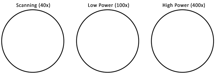
Sketch the cell at low and high power. Label the nucleus, cytoplasm, and cell membrane of a single cell. Draw your cells to scale.
Analysis
1. Why is methylene blue necessary?
2. The light microscope used in the lab is not powerful enough to view
other organelles in the cheek cell. What parts of the cell were visible.
3. List 2 organelles that were NOT visible but could be found in cells if you had a microscope with a better magnification.
4. Keeping in mind that the mouth is the first site of chemical digestion in a human. Your saliva starts the process of breaking down the food you eat. Keeping this in mind, what organelle do you think would be numerous inside the cells of your mouth?


Other Microscope Resources
Introduction to the Microscope – E Lab – explore how to use a light microscope (Letter E Slides)
Observing Plant Cells – microscope observation of onion and elodea
Comparing Plant and Animal Cells – compare onion cells to human cheek cells
Investigation: Banana Starch and Plastids – compare a ripe banana to a green banana by viewing plastids stained with iodine