
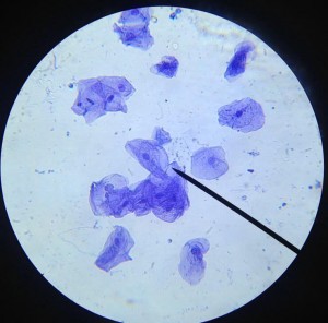
This cell activity introduces students to cell size, scale, and specialization through a hands-on microscope investigation. Students prepare and observe a slide of their own cheek cells with simple laboratory instructions. Basically, students use a toothpick to gently scrape the inside of their mouth. Then, they swirl the toothpick in a drop of methylene blue and add a coverslip. They can then view their cells with a light microscope.
When viewing the cells, students identify visible cell structures, such as the nucleus, cell membrane, and cytoplasm. In the process, they get a refresher on how to use the light microscope. Most students in an anatomy class have already completed basic biology, so they have a basic understanding of microscopes.
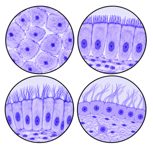
Students then compare cheek cells with different types of cells found throughout the human body. This includes epidermal, digestive, and respiratory cells. They examine differences in cell size, shape, and structure and consider how these differences relate to each cell type’s function. Though the instructions do not have them view these cells, if you have specimens, this is a great opportunity for more practice with the microscope.
The activity also introduces students to micrometers (µm), magnification, and scale bars, helping them understand the difference between how large a cell appears under the microscope and its actual size. It works well as an introduction to microscopy, cell structure, specialized cells, or size and scale in a high school biology unit.
Related Resources
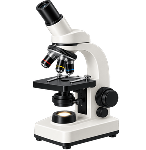
Introduction to the Microscope – E Lab – explore how to use a light microscope (Letter E Slides)
Microscope Investigation for AP Biology – a more detailed lab for advanced students
Types of Cells Coloring – color the parts of the cell and different kinds of cells found in the human body
Giant Microbe Cell Plushes – this set has most of the body cells, add them to your lecture for a visual reference!
Quiz: Parts of the Cell – Self grading quiz to test your knowledge of cells