
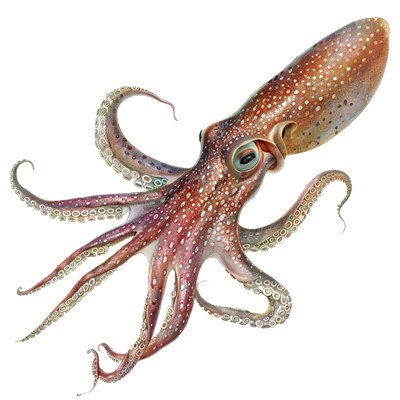
Squids, with their fascinating biology and mysterious allure, have long captivated the curiosity of scientists and students. From their unique propulsion mechanisms to their complex nervous systems, there’s much to learn by exploring the inner workings of these intelligent marine creatures.
In this dissection, you will first explore the external anatomy of the squid. The squid has many adaptations tailored for life in the ocean. They use a water jet to propel them through the water, and their streamlined shape is very efficient! Students will observe the many features of the external anatomy and internal anatomy with this comprehensive dissection guide.
What do you need? A squid specimen purchased from biological supply companies or Amazon. Optionally, you may also want to have a variety of illustrated guides to compare the specimen with drawings and diagram, like the “Illustrated Dissection Guide to the Squid.” One of my favorite lab resources for any dissections are the Concise Guides, which have full color images. You will also need dissecting tools and pans, and preferably sinks for clean-up. You can substitute a hand lens for the dissecting microscope for viewing the sucker disks.
Download the Squid Dissection Guide which includes instructions and diagrams for students to label. It is formatted for Google Doc, so you can make a copy and edit as needed for your class. This guide replaces a simpler version I use in introductory biology.
For students who are absent or choose not to participate in the dissection, you can use the virtual squid dissection as an alternative.
*Updated 2025
Part 1: External Anatomy of the Squid
First, students count the number of arms and tentacles and locate eyes and water jet. They will also need to determine the dorsal and ventral side of the animal. Generally, the dorsal side of the squid is darker in color than the ventral side. You can even discuss how countershading works to camouflage aquatic animals. Students get practice with measuring as they determine the length of the mantle in centimeters.
Once the students have determined which side is up, they will count the number of arms and the number of tentacles for their specimen. With a dissecting microscope, students take a closer look at the sucker disks located on the arms and tentacles, as well as suggest a purpose for those disks.
Next, students extract the buccal mass of the squid to observe the sharp beak found within. In removing the buccal mass, they may also observe the anterior end of the esophagus.
Once all the external structures have been found, students label a drawing and answer questions regarding their observations.
Part 2: Internal Anatomy of the Squid
Students will make a longitudinal cut on the ventral side of the squid to expose the internal organs. They will locate the stomach, esophagus, gills, hearts. They will then remove the ink sac and use the ink to scribble their initials on their lab paper. Students will also determine the sex of the squid by locating the gonad in both males and females, and the nidamental gland (only females).
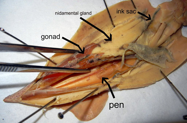
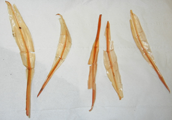
Finally, students will carefully remove the pen of the squid. The pen is a stiff structure located on the dorsal side of the squid, just underneath the mantle. The purpose of the pen is to provide stability when swimming, it acts like a little backbone, though it is not made of bone. Instead, the pen is made from chitin, which is the same material that makes up the shells of other mollusks. I make it a challenge for them to try to remove it all in one piece.
Students can also take the practice quiz over the squid to test their skills!