Virtual Squid Dissection
This page can be used as a substitute for a hands-on squid dissection. Students without access to squid, or who were absent the day of the dissection can view photos of the squid and complete the lab guide.
Related: Google Slides Virtual Squid Dissection
Step 1: Examine the External Anatomy of the Squid
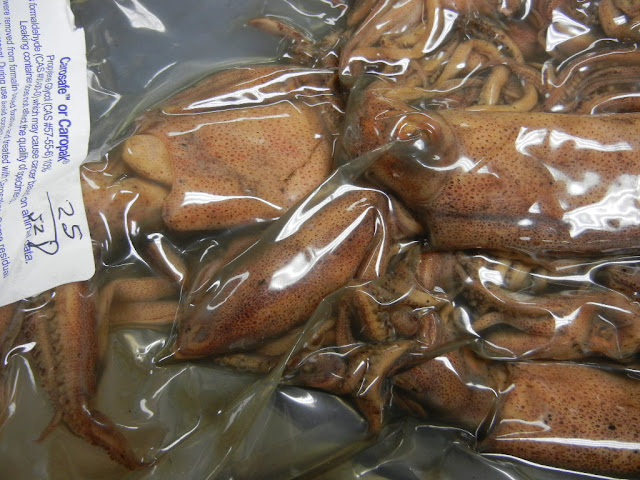
Squids are shipped in bags and are stored in a preservative, when you first open the bag, you might notice a pungent aroma. Rinse the squid off and lay it in a dissecting pan.
Determine the dorsal (back) side of the squid by looking for a darker coloration and the presence of fins.
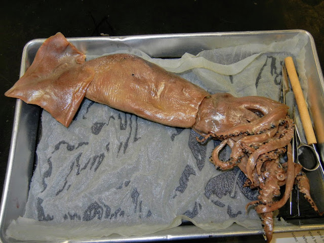
The eyes are quite large for an invertebrate and lie on either side of the head. Note the mantle that surrounds the main part of the body. The squid has two main parts: the mantle (with the fin) and the head region that also contains the tentacles (foot). In fact, that is why they are called CEPHALOPODS, the word translates to mean "head-foot."

Stretch the arms out so that you can count them and locate the two longer tentacles. How many arms are visible?
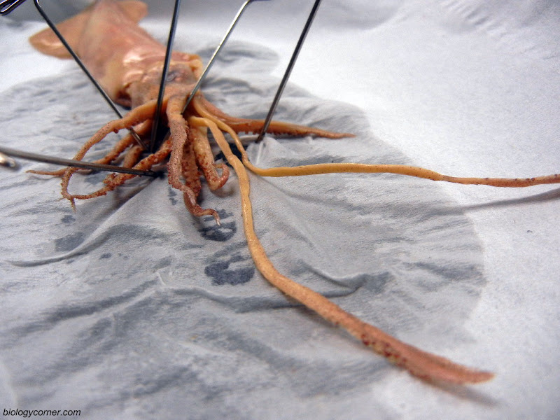
Each arm and tentacle comes equipped with suckers to help them latch onto and hold prey.

The ventral side of the squid is lighter in color and the water jet is clearly visible from this angle.
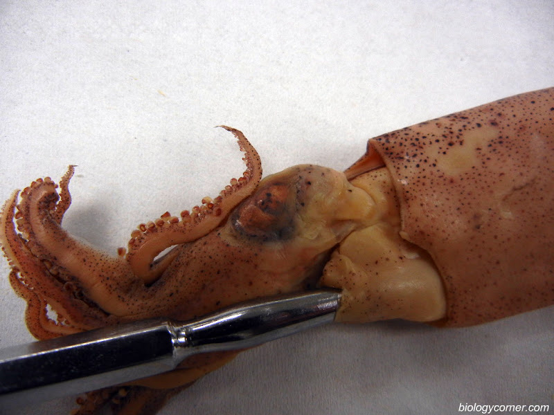
The mouth is found in the center of the arms. Pull the arms apart so that the mouth can be located.
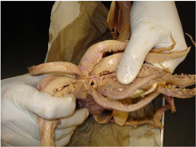
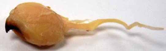
With a probe, you can feel the hard structure inside the mouth known as the beak. The beak is used to tear prey and can be very sharp (and deadly to fish!). To remove the beak, you probably will need to also remove the muscular bulb that surrounds it, known as the buccal bulb. The buccal bulb (or mass) can be removed by cutting at the base of the tentacles where the mouth is located.

In the next photo, you can see the buccal mass attached to a long tube. That tube is the esophagus. Cutting through the muscle of the buccal bulb will reveal two jaws. The jaws are also called the BEAK because it resembles the beak of a bird. This structure is sharp and strong and is used to tear prey, such as fish.
Part 2: Internal Anatomy of the Squid
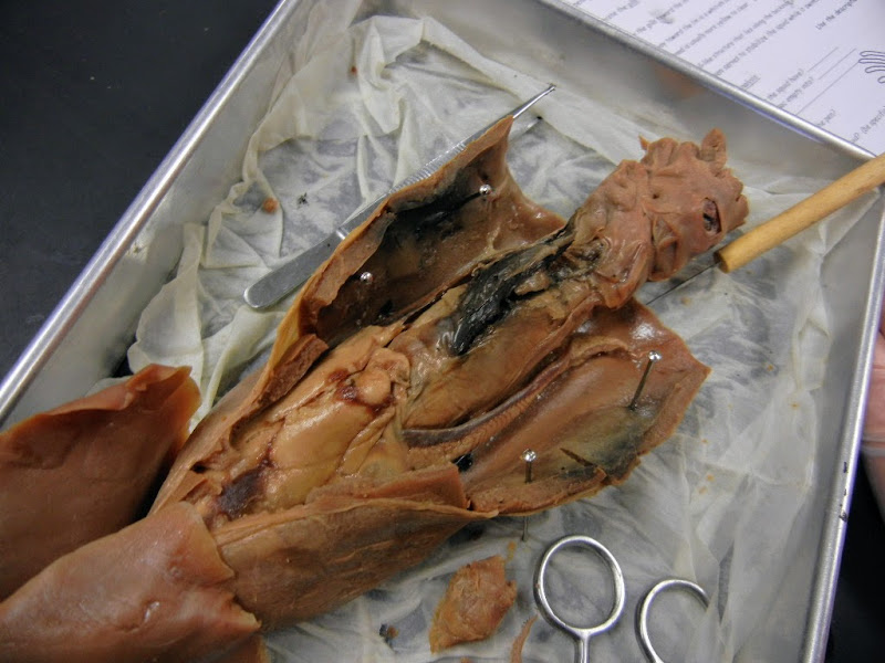
Scissors are the best tool to open the squid's body cavity. You can easily raise the mantle just where the water jet is located and cut to the anterior end of the squid (toward the fins). The photo below shows the mantle of the squid cut, but has not been pinned. The mantle is rubbery and will not stay open unless you pin it open.

Pin the mantle open so that the inside of the squid can be viewed. You may see eggs if you have a female.

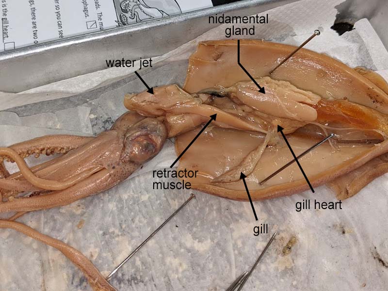
One of the more obvious structures in the squid is the ink sac. It is visible as a dark structure in the middle of the body cavity near the water jet.
The feathery structures that lie on either side of the body cavity are the gills, at the base of each you find a heart, or more accurately called the gill-heart.

The large structure in the middle of the squid is called the nidamental gland. It is more noticeable in female squids.

If you lift the nidamental gland, the stomach lies just underneath it. The stomach is often a thin membraned structure that can easily be missed among the other organs of the squid.
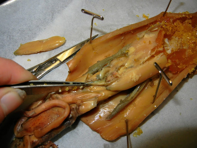
At the water jet, are long structures that are used for movement - the retractor muscles.
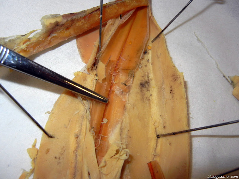
If you push all of the organs of the coelom to the side, you can locate and remove the PEN of the squid. The pen is a hard structure that is used to stabilize the squid when its swimming. If you are careful, you can remove it in one piece.
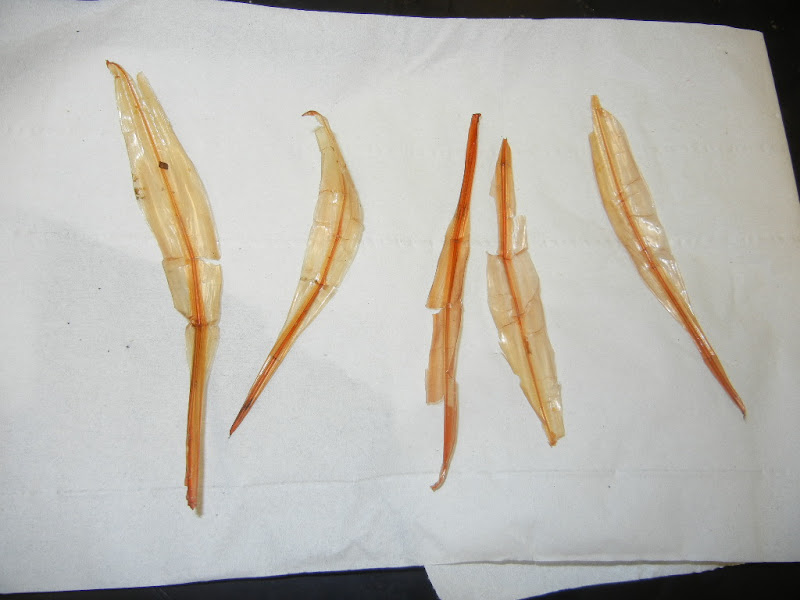
These are pens that have been successfully removed.
Part 3 - Clean Up
The squid is often a messy dissection, trays are lined with paper towels to help with the cleanup process. Since a virtual lab skips the messy part, consider a mental clean-up of the structures you need to know. Students were also required to label the squid. Print out the Squid Dissection Guide and make a sketch of the external anatomy and label the internal anatomy of the squid.
See entire squid dissection album on Google Photos
Part 4 - Label the Squid
Each of the following are found in the squid, visualize where they are located and what they look like.
Stomach
Ink sac
Esophagus
Gill
Heart
Gonad
Anus
Pen
Arms
Tentacles
Water Jet
Retractor Muscle
Nidamental Gland
Buccal Bulb