
Muscle anatomy can be a challenging topic for beginning anatomy students. The words are strange, hard to spell, and all kind of sound a like. I spend at least two days just working with them on the names and how all of the parts are arranged. Students will eventually learn how muscles contract (sliding filament theory), but that’s not until they have a good understanding of muscle structure.
In order to help my students with the anatomy of the muscle, I have several exercises to support their learning. We start with the basic connective tissue that surrounds the individual muscle fibers.
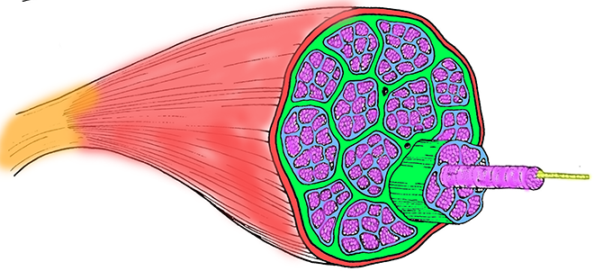
- Epimysium is the outermost layer of connective tissue that surrounds an entire muscle. It encases the entire muscle and separates it from other muscles and structures in the body.
- Perimysium is found within the muscle and surrounds bundles of muscle fibers called fascicles. Fascicles are groups of muscle fibers that are similar in function and often work together to produce specific movements.
- Endomysium is the innermost layer of connective tissue and is located within each fascicle, surrounding individual muscle fibers, also called myofibrils.
Each myofibril contains individual myofilaments. There are two types: actin (thin) and myosin (thick).
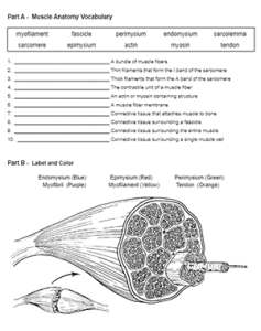
Students complete this basic worksheet: Muscle Anatomy and Vocabulary to reinforce those terms. I also have a version of this activity that can be completed online using google slides.
The worksheet begins with a list of terms and descriptions. In the second section, students color the muscle with a focus on the connective tissue and myofibrils.
Sarcomere Anatomy
Once they have a good mental model of the muscle and its fibers, we go on to talk about the other components of muscle cells. This includes the sarcoplasmic reticulum, sarcolemma, t-tubules, and sarcomeres.
A sarcomere is the basic contractile unit of a skeletal muscle. It is the structural and functional unit of a muscle fiber (muscle cell) responsible for muscle contraction. Sarcomeres are highly organized, repeating units that give skeletal muscles their striated (striped) appearance when viewed under a microscope.
There’s a coloring activity on the sarcomere too!
Finally students learn how the muscles communicate with the nervous system. They can color and label the neuromuscular junction.
Other Resources on Muscles
Labeling Muscle Structure with Flashcards