
First year biology students explore a variety of animals during dissections and live experiments. Crayfish are an inexpensive way to introduce students to invertebrate morphology. My students do a comparative anatomy where they dissect a squid, then the crayfish, and finally the frog (or fetal pig).
Students might call this animal other names, such as crawdads, crawfish, or even mudbugs! In this lab, students examine the external anatomy of a crayfish, a member of the Phylum Arthropoda.
The Crayfish Anatomy Worksheet includes instructions for locating and identifying external features of the crayfish, and then the internal features. The guide includes specific instructions and diagrams, though I recommend providing students with additional resources, like The Illustrated Guide to the Crayfish.
Overview of the Activity
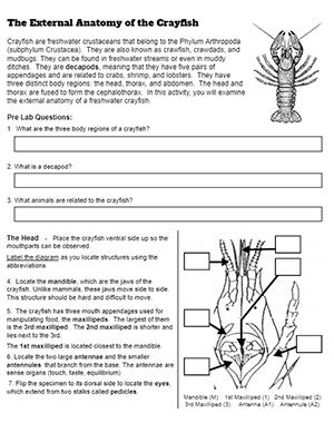
First, students look at the head and mouthparts of the crayfish. The mandible (jaws) of the crayfish open side to side. There are three maxilla which are used to manipulate food. Crayfish have two long antennae, and two shorter antennules. Once students have found the structures, they label a diagram of the head.
Next, students explore the body sections of the arthropod. Crayfish have a cephalothorax (head and thorax) covered by a carapace. The abdomen is segmented, with the final segment containing a fin-like structure called uropod.
Students then examine the appendages of the crayfish. There are four pairs of walking legs and small swimmerets attached to each segment. The largest appendage is the cheliped, or the claw. Students remove the claw and manipulate it to see how how many joints it has.
Finally, students determine whether their specimen is a male of a female. Then they measure the length of each crayfish and compare the size of males to females. Generally, males are larger than females. A short labeling and coloring exercise is included for labeling all of the structures found on the external anatomy of the animal.
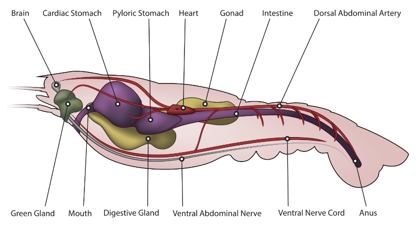
In the second part of the activity, students remove the carapace and locate the internal structures, such as the green glands, heart, stomach, and intestine.
Other Arthropod Resources
- Anatomy of the Crayfish Virtual Lab – includes photos and descriptions
- Crayfish Labeling – simple diagram with word bank
- Arthropod Coloring – compares anatomy of crustaceans, spiders, and insects
- Dating Profile for Arthropods – short research project to showcase a single species
- Arthropod Anatomy – Google Slides – includes images and diagrams to learn the anatomy