
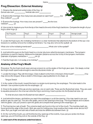
The external anatomy of the frog includes an investigation of the main features of the frog and the anatomy of the mouth. Students examine the front and hind legs, measure the lengths of frogs, and the diameter of the tympanic membrane.
Finally, students cut the angles of the jaw to reveal the vomerine and maxillary teeth, the glottis, esophagus and Eustachian tubes. A probe is used to show how the eustachian tubes connect to the tympanic membrane. Students remove the nictitating membrane of the eye and can optionally remove the lens (eyeball). Students then label an image of the frog’s head to indicate that they have located the main features.
This worksheet is part of a large unit covering frog anatomy:
Complete Frog Dissection Packet – handout for students that includes the external and internal anatomy, brain, and leg bones. Includes a list of terms to study for lab practical.
You can use any of the photos in this album for display and discussion.
Grade Level: 8-12 | Time Required: 45 – 60 min
HS-LS1-2 Develop and use a model to illustrate the hierarchical organization of interacting systems that provide specific functions within multicellular organisms