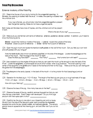
Many biology classes include a fetal pig dissection. My freshman honors biology class dissects the pig a few weeks after the dissection of the frog. In this way, students can get a detailed understanding of anatomy and how the physiology of the amphibian differs from that of a mammal.
The unit ends with a lab practical, where students identify structures on pinned pigs. The same assessment is done after the frog dissection, so students are familiar with the process. They usually do better on the pig than the frog. This is likely due to the fact that there are similar structures and familiarity with the terms.
In addition to structures of the digestive system, respiratory system, and urogenital system, students learn about the circulatory system. Double-injected fetal pigs help students visualize the path blood takes through the body.
The fetal pig dissection takes a little longer than the frog dissection, but most of the specimens will keep for at least a week.
 Student Resources
- Fetal Pig Dissection – Student Handout with directions, questions, and images
- Gallery: Fetal Pig Dissection – photos from dissection with labels
- Fetal Pig Dissection Word List – listing all the structures located during the dissection
- The Ultimate Fetal Pig Dissection Review – images and links to supplement dissection
- Virtual Pig Dissection – collection of images and descriptions from Whitmen.edu
Grade Level: 9-12
Time Required: 3-4 hours
NGSS Standards
HS-LS1-2 Develop and use a model to illustrate the hierarchical organization of interacting systems that provide specific functions within multicellular organisms
HS-LS1-2 Develop and use a model to illustrate the hierarchical organization of interacting systems that provide specific functions within multicellular organisms

