
I find that my students really have difficulty with meiosis. I’m not as concerned with them memorizing the phases (though that is part of the lesson) as I am with them understanding how the process contributes to variability.
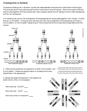
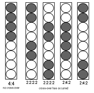
I have several worksheets the model the process, like the “meiosis label” slides where students view images, label diagrams, and answer questions. Students view how chromosomes form a tetrad and exchange DNA during prophase. They can also see how recombinants form based on how the chromosomes arrange during the two cell divisions. Senior students in AP biology usually need a refresher on the process, so I give a short slide lecture before heading to the activities.
Students often have difficulty transferring a paper model to actual real-life examples of recombination. Because this is all occurring at a microscopic level, it can’t easily be viewed with basic classroom equipment.
Luckily, there is an fungus which reveals the end product of the process, Sordaria fimicola.
Fungus Among Us
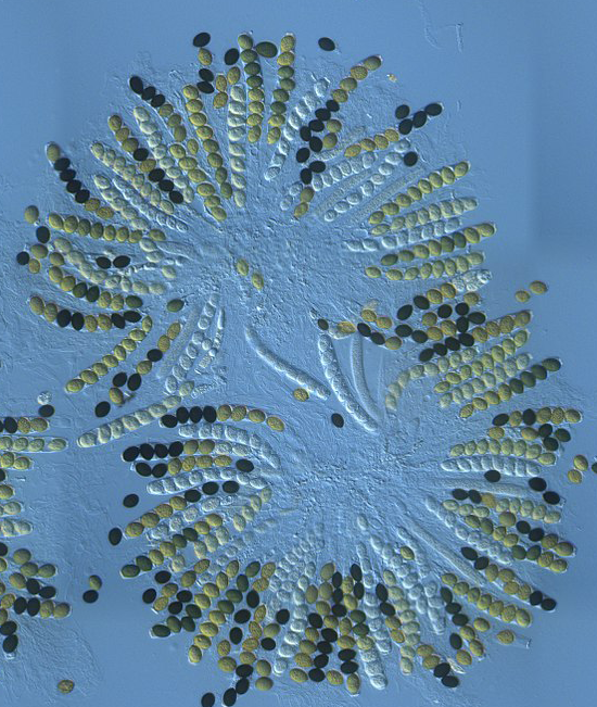
Sordaria is a fungus that is common in the feces of herbivores. The cells of the fungus are haploid, and to reproduce it combines with another haploid cell to create a diploid zygote. That zygote undergoes meiosis to produce haploid spores. The spores reveal crossing over in genes that control the color of the spore.
There are several labs using ascospore images that can be purchased, but I created this one with a photo from Wikimedia commons.
The activity includes background information about how the fungus reproduces and how to count crossover spores. Students then calculate the crossover frequency from an image to determine the distance from the centromere in map units.
You can also purchase sordaria slides so that students can view them with the microscope. This establishes an important link between the photo they are viewing and how that photo was obtained.