
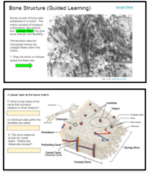
This activity was designed for remote learning where my anatomy students could learn the parts of the bone matrix. Usually, this topic is learned with manipulatives, labeling practice, and even modeling clay.
With students learning from home, I needed to get creative with how students could learn how bone is organized and how it grows and remodels.
Students view each slide and are asked to perform a task, like labeling or answering a question about the diagram shown. I find students do not always engage fully with diagram 2D models, so by asking them specific questions, students must focus their attention on the diagram and how the parts fit together.
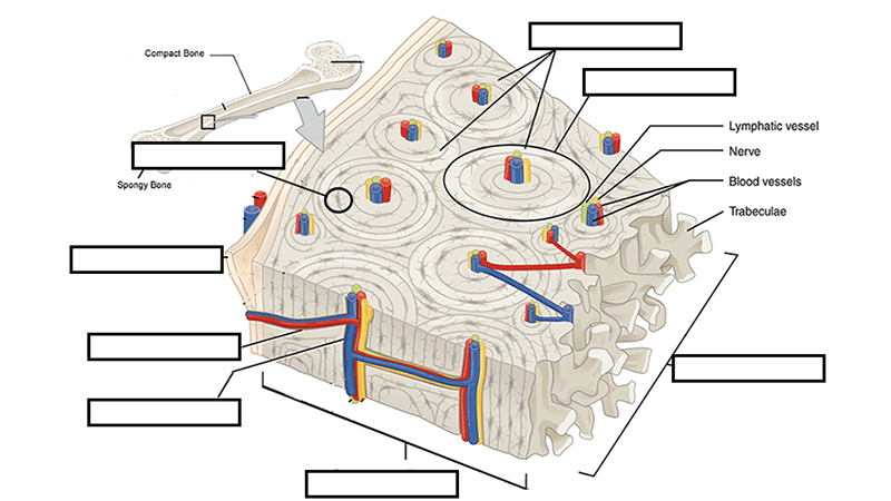
After examining the diagram students are then shown one where then need to drag and drop the labels to the image to label it. The image is the same as the ones they already viewed, and they can easily refer back to those slides if needed. On this image, students label the osteon, lamellae, central canal, osteocyte, and periosteum.
Students are also given information on how bone grows and remodels (osteoblasts and osteoclasts). Here they simply need to drag a short definition to the space on the diagram. These activities aren’t difficult, as they are intended to work as an interactive lecture, with students engaging in the material by labeling and answering questions as they go.
The final task in the set sends students to a virtual microscope where they can view bone tissue at 4x, 10x, and 100x. Here they are asked to take a screen shot of what they see under the microscope and label structures.