

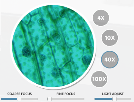
This worksheet can be used with the Virtual Microscope where students can place specimens on a stage and use coarse and fine adjustment knobs to magnify up to 100x.
Generally, I have my students practice with real microscopes, starting with a basic tutorial lab where they focus on the letter “e.” This virtual lab also starts with the letter “e” and then has students look at plant cells and human blood and make comparisons.
In the lab version, students would compare onion cells to their own cheek cells. I prefer cheek cells to blood cells because cheeks cells are much larger and have a visible nucleus, but this simulation does not have cheek cells. Blood cells are a fair substitute, but you cannot see a nucleus within them. Red blood cells don’t have nuclei.
The controls are pretty straightforward and allow students to focus and adjust lighting. When switching to 100x, students must add oil to use the oil immersion lens.
The google doc and pdf download contain two versions, one which allows for students to make drawings of the specimens at different magnifications. The other version is intended for distance learning so that students can upload the final document into an LMS without having to scan drawings. In that version, students take screenshots and add them to the document. This was included in case we aren’t able to do physical labs this year due to the danger of Covid-19.
The site hosting the virtual microscope also has downloadable pdf worksheets that can be used, the one I made is similar but designed it to be a substitute for labs I do with freshmen. This could also be used as a way for students to make up work if they missed the lab.
The Google Doc can be easily modified and shared on Google classroom. The PDF version has form fields that might be better with other LMS.