
One of the biggest challenges when teaching the muscular system is helping students understand how a nerve impulse actually leads to a muscle contracting. Terms like acetylcholine, calcium ions, troponin, tropomyosin, ATP, and myosin heads are introduced in quick succession, making it easy for students to memorize vocabulary without understanding how everything fits together.
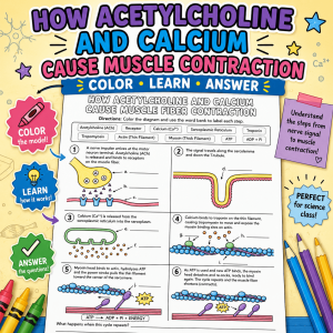
This Muscle Contraction Coloring Worksheet was designed to slow the process down and help students visualize each step of the sliding filament model.
From Nerve Signal to Muscle Movement
Instead of presenting muscle contraction as a single event, this activity walks students through the entire sequence.
Students begin at the neuromuscular junction, where a motor neuron releases acetylcholine (ACh). They follow the signal as it travels across the sarcolemma and down the T-tubules, triggering the sarcoplasmic reticulum to release calcium ions.
The worksheet then illustrates how calcium binds to troponin, causing tropomyosin to move away from the binding sites on actin. Once those sites are exposed, myosin heads attach to actin and use ATP to generate the power stroke that pulls the thin filaments toward the center of the sarcomere. As this cycle repeats, the muscle fiber shortens, producing contraction.
By seeing each step in order, students gain a much stronger understanding of how these molecules work together.
What’s Included
The resource includes:
- A detailed muscle contraction coloring worksheet
- Step-by-step illustrations of the sliding filament model
- Labeling activity
- Student comprehension questions
- Teacher answer key available at TpT
Related Resources
Muscle Anatomy Vocabulary – reinforcement for terms used in the musclular system
Case Study: The Mystery of Muscle Weakness – explore how myasthenia gravis affects the muscles and neurons
Slides and Guided Notes on the Muscular System
Understanding Muscle Structure – labeling activity that focuses on the sarcomere and microscopic structures