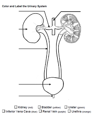
This simple worksheet asks students to label the major structures of the urinary system. They can also choose to color the diagram. I use coloring sheets in anatomy and physiology classes but this could also be used in biology or as a supplemental graphic for a frog or fetal pig dissection.
Though many of my students like coloring exercises, I do have a few that really dislike coloring. This worksheet doesn’t need to be colored and students who don’t care to color it can still benefit from the labeling. In addition to paper models of the urinary system, students are also given models to locate the structures. Anatomy students also dissect a kidney which are fairly inexpensive to purchase.
This coloring worksheet can be paired with a related activity where students label and color the internal structures of the kidney: nephrons, renal pyramids, renal pelvis.
Time Required: 10-15 minutes
Grade Level: 7-12
Download PDF Google Doc Key (TpT)
Ruth Seabaugh
Thank you so much for putting your materials on the internet so other teachers can use them as well. I think there is a problem with this diagram. It appears that this is from a posterior view (since this drawing has the ureters behind the iliac artery and vein.) Even if it is a posterior view, the aorta and vena cava appear to be drawn incorrectly at the point of the renal artery/veins. I did look at your more recent post with the online fill in the blanks urinary system and that diagram is correct. Just wanted to let you know.
Many thanks again for putting your work out there!
Admin
Thanks for letting me know! I’ll have to make some adjustments, though this is not my original drawing. I pull most of the images I use here from wiki and creative commons clipart. The line diagrams can be hard to find, or I can adjust other drawings within photoshop. I’ll see what I can do!