
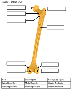
As part of the skeletal system unit, anatomy students explore each bone and their structures. For example, each group is given a femur (I have a collection of real and plastic models) and label structures. Structures include the trochanters, condyles, head, neck, and linea aspera. Students use clay and toothpick flags to label each structure and upload photos to Google classroom.
Students enjoy the process, but struggle with the final test, which is a lab practical. For this test, students go from station to station and label tagged structures on all of the bones. It is very challenging for them!
One of the reasons I do this activity every year is to prepare students for lab based classes they may encounter in college. Many have done a similar activity their freshman year with the frog dissection.
In order to help them learn the structures, I provide them with many supplemental resources. The worksheet on labeling the bones shows images of the shoulder, pelvis, legs, and arms. Students then use the word bank to label the structures. You can assign this as practice, or even as an assessment.
I created the images from Wikimedia files and added the boxes and text to the documents. During the lab, I encourage students to use their phones and lab manuals to locate the structures from the lab guide.
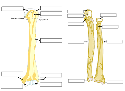
Bones from the Worksheet
I have two versions of this labeling activity. One is on paper and can be printed as a handout. The other uses the same images on Google slides that students can label by dragging text to the images of the bones.
Other Activities for Learning the Bones
- Lab Guide
- Lab Practical (Practice)
- Bone Flashcards (Printables)
- Skull Labeling