Heart - Right Atrium and Ventricle
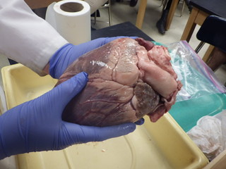
With a single cut through the right side of the heart, the right atrium and right ventricle are revealed. Preserved hearts can be somewhat rigid, you may need to hold the sides of the heart open to see the internal structures.
The right side of the heart is less muscular than the left side; this is because they only need to send blood to the lungs and back, which is a much short path than delivering blood to the entire body, which is the job of the left side of the heart.
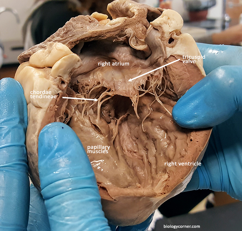
The tricuspid, or right atrioventricular valve, lies between these two chambers. The chordae tendineae anchor the tricuspid to the papillary muscle. These muscles appear smooth and slightly raised on the interior of the chamber.
The pulmonary artery connects to the right ventricle of the heart. This can be seen by placing a probe in the artery.
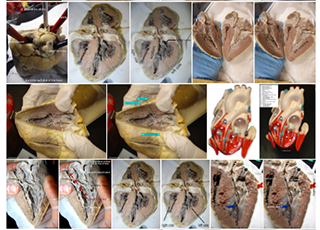
Gallery on Flickr