Heart - Left Atrium and Ventricle
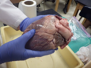
A coronal cut reveals the left atrium and left ventricle. It should be readily apparent that the left side of the heart is much thicker than the right side. This is because it must pump oxygenated blood out of the aorta and that blood must be delivered to the entire body and make the return trip. This requires a very large force.
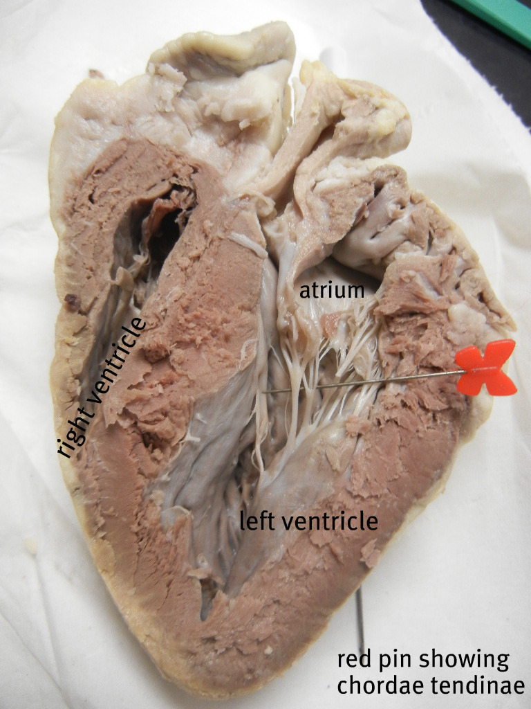
The bicuspid, or left atrioventricular valve, lies between these two chambers. The chordae tendineae anchor the tricuspid to the papillary muscle. These muscles appear smooth and slightly raised on the interior of the chamber. This valve is also called the mitral valve.
Blood returning from the lungs enters the left atrium via the pulmonary vein. From there blood travels to the left ventricle and is pushed out the aorta. This contraction of the heart is called the ventricular systole. Relaxation of the ventricle is called diastole. These two values correspond to a blood pressure reading. The typical reading is 120/80, where the higher number corresponds to the contraction of the ventricle, and the lower number is relaxation.
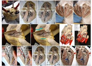
Gallery on Flickr