Learn the Anatomy of the Heart (by Number)


The human heart is similar to the hearts of other vertebrates. Mammals and birds (and some reptiles) have what is known as a double-loop circulatory system , where blood leaves the heart, goes to the lungs where it becomes oxygenated and then returns to the heart before delivering the oxygenated blood to the rest of the body. The heart has four chambers, and most diagrams will show the heart as it is viewed from the ventral side. This means that as you look at the heart, the left side refers to the "patient's" left side and not your left side.
**For each of the numbers described below, LABEL on the heart diagram.**
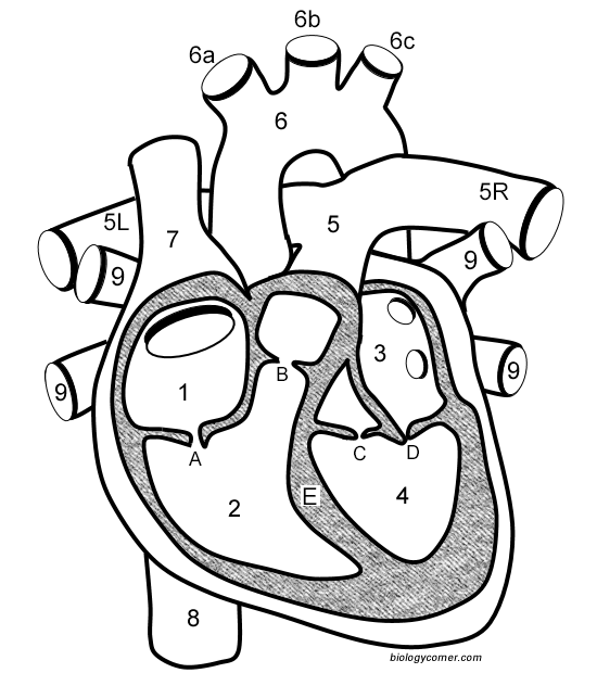
 Blood that has traveled through the body supplying nutrients to tissues eventually returns to the heart through the superior vena cava (7) and the inferior vena cava (8). Color both dark blue.
Blood then enters the right atrium (1) where a small contraction pushes blood through the tricuspid valve (A) and into the right ventricle (2). Color the right ventricle dark purple.
From the right ventricle, blood is pushed out through the pulmonary valve (B) and into the pulmonary trunk (5). This artery branches into two arteries, the left pulmonary artery (5L) and the right pulmonary artery (5R) which will deliver the blood to the lungs where it will become oxygenated. Color the pumonary trunk and arteries light blue.
Oxygenated blood returns from the lungs through the left and right pulmonary veins (9) which empties into the left atrium (3). Color the pulmonary veins pink. Color the left atrium red.
From the left atrium, blood goes through the bicuspid valve, which is also called the mitral valve (D) and enters the most muscular part of the heart, the left ventricle (4). A powerful contraction of the left ventricle will send blood through the aortic valve (C) and into the largest artery of the body, the aorta (6). Color the left ventricle orange. Color the aorta dark red.
Blood enters the aorta and will travel to the head and shoulders through three smaller arteries: the brachiocephalic (6a), the left common carotid (6b), and the left subclavian (6c). The aorta forms an arch as blood is routed to the lower part of the body where it oxygenates organs and muscles. Color dark red.
Red the descriptions carefully and trace the flow of bloon in the heart using arrows.
Don't forget to LABEL the parts of the heart on the diagram!
1. Compare the location of the tricuspid and bicuspid.
2. Compare the direction of blood flow in the pulmonary artery to the pulmonary vein.
3. Mitral regurgitation is a heart condition that occurs when the mitral valve does not close fully. Based on your knowledge of the heart, describe what happens to the blood of someone who has this condition.
4. A ventricular septal defect occurs in some newborns where the septum (E) does not close completely during development. Consider a ventricular contraction where there is a hole in the septum. Blood that would normally travel out the aorta would go where?