 Rat External Anatomy


Procedure: Obtained your rat. Rinse it off with water and place it in your dissecting pan to observe the general characteristics. Make sure you know each of the highlighted words. The rat's body is divided into six anatomical regions:
cranial region - head
cervical region - neck
pectoral region - area where front legs attach
thoracic region - chest area
abdomen - belly
pelvic region - area where the back legs attach
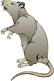
1. Note the hairy coat that covers the rat and the sensory hairs (whiskers) located on the rat's face, called vibrissae. ![]()
2. The mouth has a large cleft in the upper lip which exposes large front incisors. Rats are gnawing mammals, and these incisors will continue to grow for as long as the rat lives. ![]()
3. Note the eyes with the large pupil and the nictitating membrane found at the inside corner of the eye. This membrane can be drawn across the eye for protection. The eyelids are similar to those found in humans. ![]()
4. The ears are composed of the external part, called the pinna, and the auditory meatus, the ear canal. ![]()
5. Locate the teats on the ventral surface of the rat. Check a rat of another sex and determine whether both sexes have teats. ![]()
6. Examine the tail, the tails of rats do not have hair. Though some rodents, like gerbils, have hair on their tails. ![]()
7. Locate the anus, which is ventral to the base of the tale. ![]()
8. On female rats, just posterior to the last pair of teats, you will find the urinary aperture and behind that the vaginal orifice which is in a small depression called the vulva. ![]()
9. On males, you will find a large pair of of scrotal sacs which contain testes. Just anterior to the scrotal sacs is the
prepuce, which is a bulge of skin surrounding the penis. The end of the penis has a urogenital orifice, where both urine and sperm exit. ![]()
 The Muscular and Skeletal System of the Rat
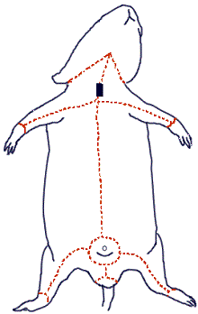
Procedure: Skinning the Rat
You will carefully remove the skin of the rat to expose the muscles below. This task is best accomplished with scissors and forceps where the skin is gently lifted and snipped away from the muscles. You can start at the incision point where the latex was injected and continue toward the tail. Use the lines on the diagram to cut a similar pattern, avoiding the genital area. Gently peel the skin from the muscles, using scissors and a probe to tease away muscles that stick to the skin.
Muscles are attached to bones by connective tissue called tendons that adhere to spines, knobs, and ridges on bones. You will need to refer to the rat skeleton to determine where the muscles are attached to bones. The end attached to the bone that does not move during contraction is called the origin. The end of the muscle that attaches to the bone that does move is called the insertion. The movement caused by the contraction of the muscle is called the action. Muscles can be easily identified from one another by their shape and overlap.
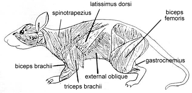
Identify the following muscles:
1.
Biceps brachii - located on the anterior surface of the humerus. ![]()
2. Triceps brachii - located on the sides and back
of the upper arm. ![]()
3. Spinotrapezius - located
across the dorsal thoracic region of the rat. ![]()
4. Latissimus dorsi - located posterior (and partially covered) by the
spinotrapezius. ![]()
5. Biceps femoris - located
on the side of the thigh, in two bundles ![]()
6.
Tibialis Anterior - located on the front of the leg. ![]()
7.
Gastrocnemius - located on lower leg, bulk of the calf muscle. Attaches
to heel by the Achilles Tendon.
8. External
Oblique - located on the sides of the abdomen.![]()
9. Gluteus Maximus - located on the lower back and rear. ![]()
10. Pectoralis Major/Minor - located in chest
![]()
Pin the muscles listed above on a skinned rat.
🐭Procedure:
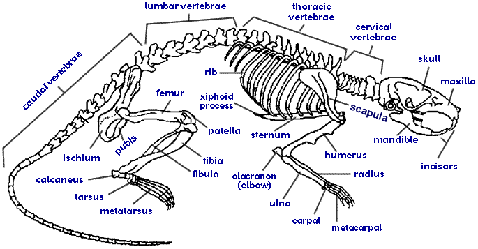
Carefully
tease away the biceps femoris and gastrocnemius to expose the 3 leg bones: Tibia,
Fibula, and Femur and the small patella (kneecap). You can
also see the ligaments around the knee that attach the bones of the lower
leg to the femur and the achilles tendon which attaches the the gastrocnemius
to the ankle.
Note that the joint of the hip is called a ball and socket
Related Resources
Rat Pages:
Rat Introduction and Guide | Rat External Anatomy
Head and Abdominal Region | Circulatory System | Urogenital System
Other Resources: Virtual Rat Dissection | Rat Dissection with Photo Submission
Coloring: Comparing a Human to a Rat Skeleton