
Investigation: How Can a Microscope Be Used to Make Observations?


Part 1: What are the Different Types of Microscopes?
Objectives:
- Compare and contrast different types of microscopes
- Learn how to identify the parts of the microscope and confidently use the microscope
- Learn the proper use of a microscope and be able to focus using each of the lenses
- Create a wet mount and stain a slide for easier viewing
- Learn how to estimate the size of microscopic objects
One of the most important tools that a biologist uses is the microscope. In this course, you will use two types of light microscopes: the compound light microscope with magnifications ranging from 40x to 400x and the stereomicroscope with magnifications ranging from 7x to 20x. Each of these microscopes focuses light through two lenses in order to magnify objects in the viewing field. Stereomicroscopes are sometimes called dissecting microscopes because they allow large specimens to be viewed closely.
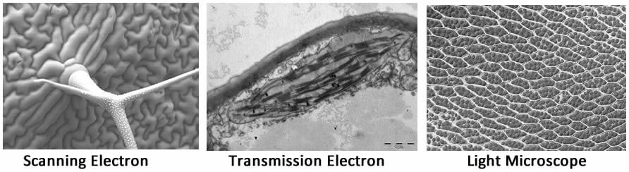
In addition to those microscopes, biologists can also use scanning electron microscopes (SEM) and transmission electron microscopes (TEM). These tools use electrons instead of light create an image of the specimen. Generally, an SEM focuses on the surface of the specimen, creating a 3-dimension view.
The TEM beams the electrons through the specimen, revealing internal structures. The electron microscopes have greater magnifications than the light microscopes, but cannot be used to view living specimens. Most beginning students will not have access to electron microscopes.
Each type of microscope has advantages and disadvantages for viewing specimens and each creates unique views. The image below shows plants as seen under three types of microscopes. The SEM shows the surface of the leaf with a close-up of a trichome, the TEM shows the chloroplast of a leaf cell, and the light microscope shows the individual cells within the leaf. A biologist will choose a microscope based on what observations she wishes to make.
Each microscope can be defined by its magnification and its resolving power (resolution.) Resolution refers to the microscope's ability to distinguish two closely spaced objects. The resolving power of the human eye is about 0.1 millimeters, meaning that small objects that are closer than that will be perceived by the eye as a single object.
1. Which type(s) of microscope would be used to:
a) View very small internal cell structures, like a chloroplast: _____________________________________
b) Look at a pond microorganisms as it swims: ________________________________
c) View the surface of a leaf to identify the number of trichomes: ____________________________
d) View the stomach of a grasshopper: _______________________________________
2. Compare the images produced by a SEM to those produced by a TEM.
3. What is the difference between magnification and resolving power?
Part 2: Getting to Know Your Microscope
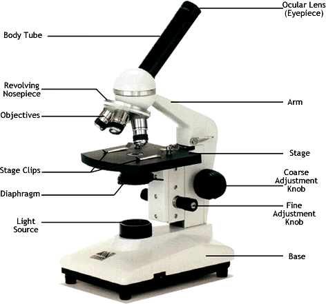
Review the parts of the microscope as they are illustrated below. You may have used a microscope in the past and have some familiarity. No matter what your starting level may be, you share a common goal with others in the class; to concentrate on improving your microscope skills. Your microscope may vary slightly from the image, but most compound light microscopes have the same general design.
A. How to Microscope Care
- Always carry the microscope with one hand on the base and the other on the arm.
- Use lens paper to clean the lenses, other types of paper can scratch the glass.
- When the high-power objective is in place, do not use the coarse focus knob.
- Do not search for objects on high power, switch to low power to find the object of interest, do not use stage clips
- Keep the microscope 3 to 6 inches from the edge of your lab table to prevent accidents.
B. What is Your Microscope's Magnification?
1. The magnification written on the ocular lens (eyepiece) is ___ (ignore any mm measurements, look for number before the X)
2. The magnification on the
Scanning objective _______ Low Power Objective _______ High Power Objective _______
3. What is the total magnification for each lens? (multiply scanning x the objective)
Scanning __________ Low Power____________ High Power ____________
C. How Can You Adjust the Light
Examine the diaphragm, determine how to change the amount of light that passes through your viewing field. Diaphragms on microscopes can be different depending on the model. Some may be shaped like wheels with several settings.
4. Describe how your diaphragm works.
D. How Can You Focus the Microscope Using the E slide?
→ Adjust the nosepiece of the microscope so that the low power (scanning) lens (4x) is in place. Place the e-slide on the stage so that you can visible see the letter "e" is over the opening where light will enter.
→ While looking into the ocular lens, use the coarse adjustment knob to bring the "e' into focus.
→ Carefully re-center the e on the slide and switch to the medium power objective (10x). The specimen will be out of focus and it may have shifted slightly. Use the coarse adjustment knob again to bring the "e" back into focus.
→ Carefully re-center the "e" again and switch to the high power objective (40x).
At this point, DO NOT USE the coarse focus knob!!
→ Use the fine adjustment knob only to bring the specimen back into focus. At this point, you will not see the letter "e," you will only see the edge, or the ink.
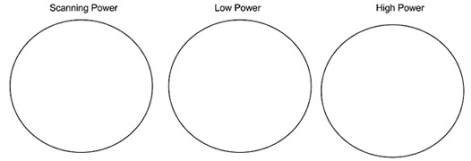
5. Draw the E exactly as it appears in your viewing field for each magnification. The circles below represent your viewing field. The E should take up as much space in the drawing as it does in your viewing field while you're looking at it.
6. Focus your e slide at each magnification. What happens to the working distance as you move from low to medium to high power? Using this information, explain why you should NOT use the coarse adjust when using the high power objective.
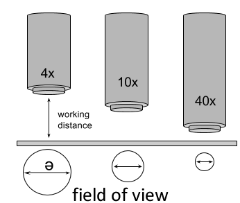
E. What is the Field of View and How Can It Be Measured?
When in focus, the distance between the slide and the tip of the objective is the working distance. The circle of light seen thought the eyepiece is the field of view. The field of view varies by microscope and by the size of the lens, a number that is usually written on the side of the lens. You will be measuring the field of view for your own microscope, which may be different than another microscope in the room.
→Use a ruler to determine the width of the viewing field under the scanning objective. Position the ruler so that the millimeter marks are visible in your viewing field, and line up one of the vertical lines so that it is just visible at the extreme left side.
→Count the number of millimeters; you may need to estimate the fraction of millimeter involved in the last section. Repeat this procedure to estimate the medium power field of view. There are 1000 micrometers in a millimeter.
7. What is your estimate of the length (diameter) of your viewing field in millimeters:
Scanning (40x)_______ Low power (100x) ________
What is your estimate of the length (diameter) of your viewing field in micrometers:
Scanning (40x)_______ Low power (100x) ________
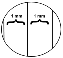
8. You cannot use this method to determine the diameter under high power (try switching objectives, you won't be able to even see the ruler). Instead you can use a mathematical proportion method to determine the diameter under high power. Show your work below with your estimate of the length (in micrometers) of your high power field of view. You are solving for "High Power Field of View."
![]()
High Power Field of View = _________
9. Sketch a paramecium and a cork slide at 400x, drawing each to scale with attention to detail.
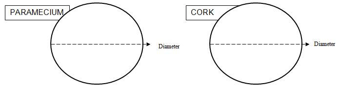
Length of paramecium = ____________ Length & Width of a single cork cell = ____________
Part 3: Working with Specimens
A. Making a Wet Mount of a Slide
→Place a drop of water on the slide. Gather a few strands of cotton from a cotton ball using forceps and place them on the drop.
→Place the coverslip at about a 45-degree angle with one edge touching the water drop and then gently let go.
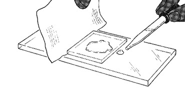
Performed correctly the cover slip will perfectly fall over the specimen and will not have air bubbles. If the cover slip does not sit flat, then you have too much fiber on the slide. If this is the case, start over.
13. Draw the specimen as it appears in your viewing field under scanning, low and high power.
B. Staining a Specimen
→ Biologists learn to stain specimens that have already been created. You will not remake this slide, but use a procedure to draw a stain under the coverslip using the capillary action of water.
→Place one drop of stain (methylene blue or iodine) on the edge of the coverslip you made with the cotton.
→ Place the flat edge of a piece of paper towel on the opposite side of the coverslip. The paper towel will draw the water out from under the coverslip, and the cohesion of water will draw the stain under the slide.
14. View the slide again, describe the appearance of the cotton fibers now compared to the view without the stain.
C. Large Specimens and Dissecting Scopes
Light microscopes are only useful for viewing small thin specimens. Stereoscopes present a larger field of viewing and handle depth much better than the light microscope. The drawback of the stereoscope is that it does not have a high magnification. Examine one of the stereoscopes in the room which will have a variety of specimens to view.
Name/Description of specimen(s) viewed: ____________________________________________________
What is the magnification(s) of the stereoscope? _____________________
Use a ruler to determine the field of view at its lowest and highest magnification:
Low Power FOV _______ High Power FOV ________
Other Microscope Resources
Introduction to the Microscope – E Lab – explore how to use a light microscope (Letter E Slides)