Investigation: Exploring Cells, a Historical Perspective


Overview: In this lab, you will explore the appearance of cells as historical figures may have seen them. Review the steps of using a microscope and focusing on high power before proceeding.
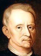
1. Robert Hooke Observed Cork
Robert Hooke was the first person to see cells when he viewed a piece of cork with a microscope. What he saw, he described as “porous” and like a “honeycomb.” From his observation he decided to call the boxes he viewed CELLS because it reminded him of the rooms that monks live in. We also use the term “cell” today to describe rooms in which prisoners live.
Find a prepared slide labeled “cork.” You should be able to see a pink specimen on the slide even before you look at it with the microscope. The cork was stained pink so that you could see it more clearly. Focus slide under scanning, then low, and then high power.
Draw the cells “to scale.” This means that you should use the circle of the viewing field to show the cells as they APPEAR when viewed at that magnification.
CORK CELLS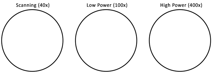
Discussion:
1) Describe what you see under the microscope using your own words.
2) How did the appearance of the cork inspire the naming of the cells?
2. Anton van Leeuwenhoek Discovered “Animalcules”
Anton Leewenhoek looked at pond water under a microscope and found that it was full of tiny like organisms that swam around. No one had a name for these organisms, so he called them “Animalcules.”
Sometimes, these organisms can swim fast and be difficult to observe. To make it easier to see one, biologists presesrve organisms on slides in a fixed state; locate a preserved slide called “paramecium.” This is an example of one of Leeuwenhoek’s animalcules, though we now know that there were also small invertebrates in the water, like the daphnia shown..
Draw the paramecium to scale.
Discussion:
3) Why are living “animalcules” difficult to observe?
4) What was Leeuwenhoek actually seeing in his sample of water (there’s no such thing as an animalcule)?
5) Do you think you could find these organisms in your drinking water? Why or why not?
3. Matthias Schleiden Loved His plants
Matthias Schleiden was a botanist that examined a large number of plants under the microscope. He concluded that all plants were made of cells, which contributed to the creation of the Cell Theory. Plants usually have square shaped cells, and the cell sof the leaves will have green chloroplasts which are used for photosynthesis. Root cells, like those of the onion, do not contain chloroplasts.
→ View prepared slides of onion cells and spirogyra under low power.
| Spirogyra (100x) | Onion Cells (100x) |
 |
|
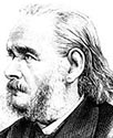
4. Theodor Schwann Loved His Animals

Theodor Schwann was a zoologist, and he spent his time looking at animal tissues with a microscope. His conclusion was similar to Schleiden’s: all animals were made of cells. Together, with other observations of the time, the CELL THEORY was developed. To see an animal cell, you will view a prepared slide of blood and a prepared slide of muscle cells. Sketch both under high power (400x)
| Blood Cells (400x) | Muscle Cells (400x) |
|
|
Discussion:
6) How was Schwann's conclusion similar to Schleiden?
7) How were plant and animal cells similar? How were they different?
Related Resources on Cells
Introduction to the Microscope – E Lab – explore how to use a light microscope (Letter E Slides)
Cheek Cell Lab – observe cheek cells under the microscope
Observing Plant Cells – microscope observation of onion and elodea