The Genetics of Sickle Cell Anemia


I. What is Sickle Cell Anemia?
A gene is a segment of DNA that codes for a protein or a trait.. Genes can be any length and sometimes involve multiple sections of DNA. The HBB gene provides instructions for making a protein called Beta-globin which is part of a large protein called hemoglobin that is found in red blood cells. Each hemoglobin protein can carry four molecules of oxygen, which is delivered to the body's organs and tissues.
If a person doesn't have enough red blood cells or the cells don't work properly, organs can become deprived of oxygen. This condition is called anemia. A person with anemia may feel tired all the time, experience difficulty with breathing, leg cramps, and dizziness.
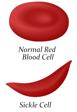
There is one type of anemia that is related to the shape of the HBB protein. When a person has sickle cell anemia, the hemoglobin protein forms long chains that change the shape of the red blood cell. Instead of a disc shaped structure that moves easily through blood vessels, sickled blood cells are shaped like bananas. The reason they have a sickled shape is because the underlying gene has the wrong instructions. These misshapen blood cells get clogged in vessels and don't have the life expectancy of normal blood cells. A person with sickle cell disease will experience fatigue (feeling tired) and have episodes of extreme pain, called a pain crisis. Sickled blood cells that block vessels in the brain can even cause stroke.
Sickle cell anemia is a life threatening disease that affects about 100,000 Americans. It is an inherited disease that is passed from parents to their children, but parents can be carriers of the gene and not have any symptoms. If both parents are carriers, their children have a 25% chance of having sickle cell anemia.
1. What is a gene?
2. What is hemoglobin?
3. How is a sickled blood cell different from a normal one?
4. Why are the blood cells the wrong shape?
5. What are the symptoms of sickle cell anemia?
6. What is a carrier?
II. How DNA Makes Protein
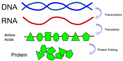
Recall that DNA contains four bases: Adenine, Guanine, Cytosine, and Thymine. The sequence of A's, T's, G's, and C's are what determines the protein that is built. Each set of three bases will code for a single amino acid. Proteins are simply chains of amino acids. To make proteins, DNA must send its code sequence to the ribosomes in the cell, but it needs a messenger to do that. Transcription is the process where DNA is converted to a molecule of messenger RNA (mRNA). The mRNA is then used to build a protein like hemoglobin.
CODON CHART
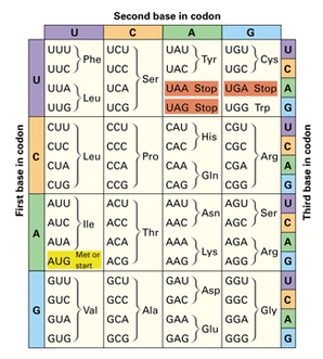
To determine the amino acid sequence of the gene, you must transcribe the DNA to RNA. The base pair rule is used to create RNA, but RNA does not contain thymine, it contains URACIL instead. This is why codon charts have U's in them and no T's.
A codon chart tells you what bases in RNA code for what amino acids. The ribosome combines all the amino acids to create a single protein, like hemoglobin. It takes three bases to determine one amino acid. Amino acids are usually abbreviated. GUC makes the amino acid valine, abbreviated as "Val" on the chart.
Here is how the codon chart could be used to determine the amino acid sequence:
DNA: A A T C A G → (DNA sequence of gene)
RNA: U U A G U C → (transcribed from RNA; follow base pair rule, no T's)
Amino Acids: Leu Val → (find on codon chart)
7. In order to make a protein, the message on DNA must be converted to what? ___________
8. How many bases in DNA are needed to code for a single amino acid? _________
9. What is a protein? _________________________________________________
10. . What base is found in RNA, but not DNA? ________
11. Consider the sequence shown, determine the complementary RNA and the amino acids
| DNA | T A C | G T A | T T T | G C A | C A C |
| RNA | |||||
| Amino Acids |
III. A Change in DNA Can Change the Protein
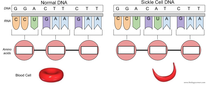
Sometimes, one of the letters in DNA gets switched with another letter, causing a mutation in the DNA. Many mutations don't have any effects, but some will change the amino acid made by the ribosomes. In the case of sickle cell anemia, just a single letter change alters the shape of the hemoglobin protein.
12. Use the codon chart to determine the amino acids created from each DNA.
13. What codon in the sickle cell DNA is altered? ___________________ (1st, 2nd, or 3rd)
14. What happens in people that have this difference in their DNA? ____________________
15. Explain how it would be possible to have a change in a single base of DNA, but have the protein NOT change and be functional. Hint: look at the codon chart.
____________________________________________________________________
TED-Ed Video on Sickle Cell (~5 minutes)
Other Resources on DNA and Proteins
Regulatory Switches in the Stickleback - based on an HHMI activity, explores gene expression in fish (Also available as a slide activity)
Gene Regulation in Prokaryotes - focuses on the lac operon and how some genes are repressible
Transcription & Translation Coloring - shows structures involved, nucleotides, base pair rules, amino acids
How DNA Controls the Workings of a Cell - examine a DNA sequence, transcribe and translate
DNA Concept Map - design a concept map showcasing the structure and function of DNA