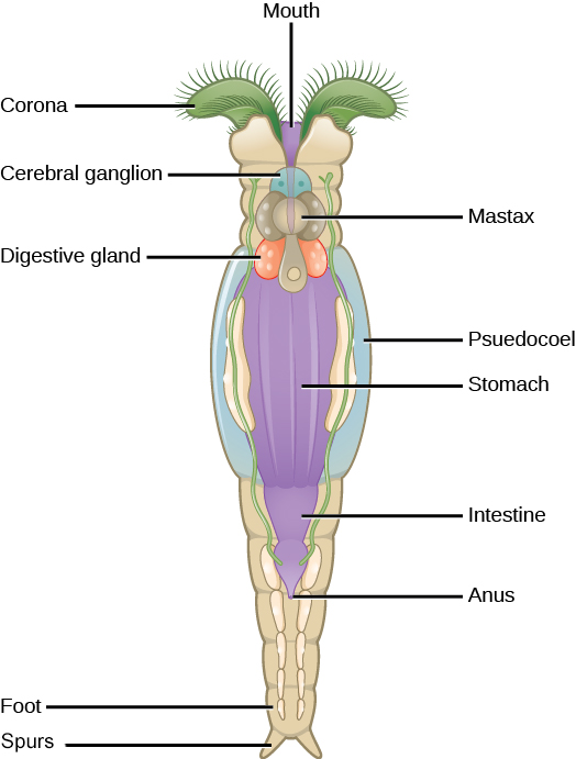
Observe Rotifers with a Microscope
Rotifers are multicelled animals. Because they are so small most people have never heard of their existence. They are about the same size as the larger unicellular organisms. They don't have a lot of cells, less than 1000, but they have some very special attributes. They are wonders of miniature design.
At the front of the body they possess a crown of hair-like cilia. They locomote by using the crown of cilia (the corona) to propel themselves. Some species walk with head and foot. Their foot (also called spurs) can secrete a sticky substance that enables them to attach to a surface.
They also use the crown of cilia to wave food into their mouth. There the food is passed into the mastax'where it is ground up and then directed towards the gut. You can often see the mastax moving as it begins the digestion process. Rotifers are so transparent that all these organs can be observed easily. They have one or two light sensitive red eye spots.
Because many species make so-called resting spores which are easily carried by the wind, they can be found anywhere if there is a little bit of water. Even in a roof gutter or in birdbaths.
Observe the Rotifer
-- Use the scanning lens to find a rotifer, they are usually easy to spot attached to algae. Some may be seen swimming across your viewing field. Switch to the LOW power objective.
1. How does the rotifer attach itself to algae?
2. What is the function of the mastax?
3. Describe how the mastax and cilia moves. What is the function of the cilia near the opening of the mouth?
4. Sketch your rotifer. LABEL (either on your drawing or the drawing above) the mastax , spurs, mouth, and gut.
Rotifer Video
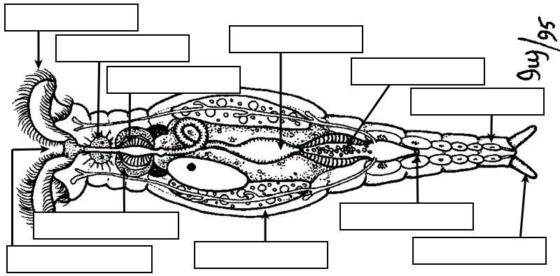
Label the Rotifer
Related Resources
Microscope E Lab - how to use the light miscroscope
Microscope Coloring - article with questions and coloring
Reinforcement: Cell Structures | Functions of Organelles
Simple Worms Lab - observe nematodes and flatworms