Student Guide to the Frog Dissection


Frog Dissection Kit from Amazon
 Dissection Instructions
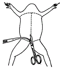
1. Place the frog in the dissecting pan.
2. Use scissors to lift the abdominal muscles away from the body cavity.
Cut along the midline of the body to the forelimbs.
3.
Make transverse (horizontal) cuts near the arms and legs.
4.
Life the flaps of the body wall and pin back.
*If your specimen is a female, the body may be filled with eggs. You may need to remove these eggs to view the organs.
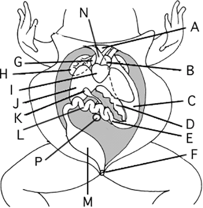
Locate each of the organs below. Check the box to indicate that you found the organs.
1. Fat Bodies --Spaghetti shaped structures that have a bright orange or yellow color, if you have a particularly fat frog, these fat bodies may need to be removed to see the other structures. Usually they are located just on the inside of the abdominal wall.
2. Peritoneum A spider-web like membrane that covers many of the organs, most easily seen covering the heart.
3. Liver--The largest structure of the the body cavity. This brown colored organ is composed of three lobes. The right lobe, the left anterior lobe, and the left posterior lobe. The live secretes a digestive juice called bile which is needed for the proper digestion of fats.
4. Heart - at the top of the liver, the heart is a triangular structure. The left and right atrium can be found at the top of the heart. A single ventricle located at the bottom of the heart. The large vessel extending out from the heart is the conus arteriosis.
5. Lungs - Locate the two spongy lungs by looking behind the heart and liver.
6. Gall Bladder --Lift the lobes of the liver, there will be a small green sac under the liver. This is the gall bladder, which stores bile. (hint: it kind of looks like a booger)
7. Stomach--Curving from underneath the liver is the stomach. The stomach is the first major site of chemical digestion. Frogs swallow their meals whole. Follow the stomach to where it turns into the small intestine. The pyloric sphincter valve regulates the exit of digested food from the stomach to the small intestine.
8. Pancreas - This structure is located on the inside curve of the stomach. It it a gland that often falls apart during the preserving process so it many not be visible on your frog.
9. Small Intestine--Leading from the stomach. The first straight portion of the small intestine is called the duodenum, the curled portion is the ileum. The ileum is held together by a membrane called the mesentery. Note the blood vessels running through the mesentery, they will carry absorbed nutrients away from the intestine. Absorption of digested nutrients occurs in the small intestine.
10. Large Intestine--As you follow the small intestine down, it will widen into the large intestine. The large intestine leads to the cloaca, which is the last stop before solid wastes, sperm, eggs, and urine exit the frog's body. (The word "cloaca" means sewer.) The opening to the outside of the doy is the anus.
11. Spleen--Return to the folds of the mesentery, this dark red spherical object serves as a holding area for blood.
12. Esophagus--Return to the stomach and follow it upward, where it gets smaller is the beginning of the esophagus. The esophagus is the tube that leads from the frogs mouth to the stomach. Open the frogs mouth and find the esophagus, poke your probe into it and see where it leads.
Removal of the Stomach: Cut the stomach out of the frog and open it up. You may find what remains of the frog's last meal in there. Look at the texture of the stomach on the inside.
What did you find in the stomach?
Measuring the Small intestine: Remove the small intestine from the body cavity and carefully separate the mesentery from it. Stretch the small intestine out and measure it. Now measure your frog. Record the measurements below in centimeters. Frog length: _______ cm Intestine length ________ cm
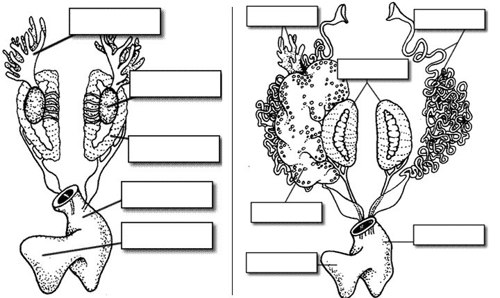
Urogenital System
The frog's reproductive and excretory system is combined into one system called the urogenital system. You will need to know the structures for both the male and female frog
Kidneys - flattened bean shaped organs located at the lower back of the frog, near the spine. They are often a dark color. The kidneys filter wastes from the blood. Often the top of the kidneys have yellowish stringy fat bodies attached.
Testes - in male frogs, these organs are located at the top of the kidneys, they are pale colored and roundish.
Oviducts - females do not have testes, though you may see a curly structure around the outside of the kidney, these are the oviducts. Oviducts are where eggs are produced. Males can have structures that look similar, but serve no actual purpose. In males, they are called vestigial oviducts.
Bladder - An empty sac located at the lowest part of the body cavity. The bladder stores urine.
Cloaca - mentioned again as part of the urogenital system - urine, sperm and eggs exit here.
Label the parts of the urogenital system.
Post Lab Questions
1. The membrane holds the coils of the small intestine together:
2.This organ is found under the liver, it stores bile:
3. Name the 3 lobes of the liver:
4. The organ that is the first major site of chemical digestion:
5. Eggs, sperm, urine and wastes all empty into this structure:
6. The small intestine leads to the:
7. The esophagus leads to the:
8. Yellowish structures that serve as an energy reserve:
9. The first part of the small intestine(straight part):
10. After food passes through the stomach it enters the:
11. A spiderweb like membrane that covers the organs:
12. Regulates the exit of partially digested food from the stomach:
13. The digestive system ends at the opening called the:
14. Organ found within the mesentery that stores blood:
15. The largest organ in the body cavity:
Label the Diagram
Credits: Title Frog at Top - http://www.clker.com/clipart-coolred-eyed-frog.html
Frog Urogenital Systems were modifed from http://biodidac.bio.uottawa.ca/
Pictures of Dissected from with labels are available at
Other Resources on the Frog
Frog Brain and Bones – remove the frog’s brain, expose the bones of the lower leg
Frog Dissection Crossword – review terms and procedures
Frog Dissection Alternative – for students who do not wish to dissect a frog, project using internet resources
Frog Anatomy Review – resource site for virtual frogs and practice quizzes
Frog Anatomy Labeling – basic pictures of frogs for students to label, serves as a review for the lab test
Frog Dissection Gallery - photos of the frog with labeled structures
Frog Dissection Kit from Amazon