Frog Dissection Coloring 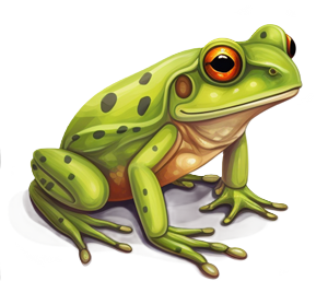
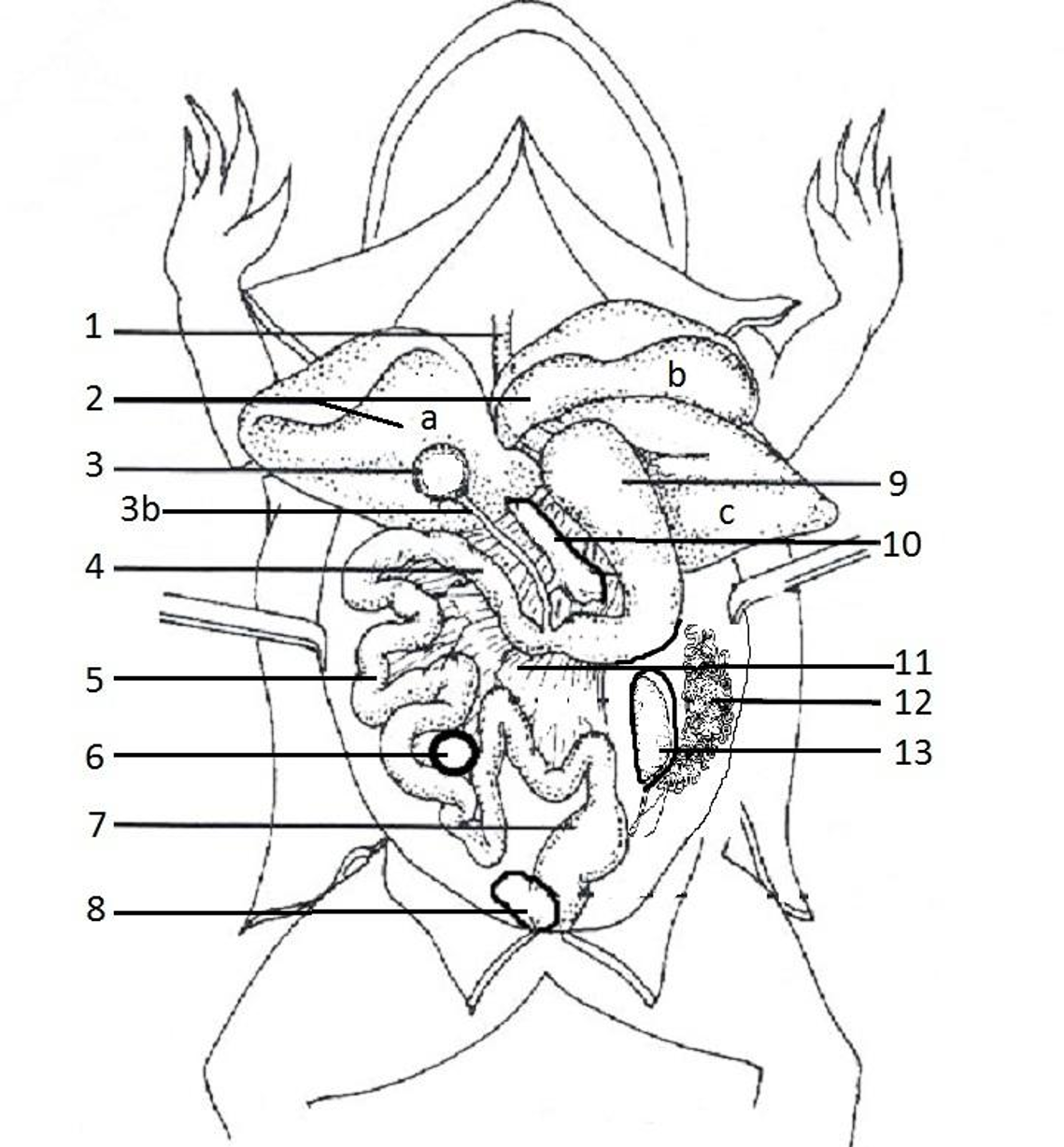
When the abdominal cavity of the frog is opened, many organs of the digestive and urogenital systems can be observed. As you read the descriptions of the organs below, color them on the diagram.
1. Leading from the mouth is a tube that connects to the stomach. Color the esophagus pink. 
2. The largest organ is the liver, and it consists of multiple lobes. Color the right lobe (a) light brown. Color the left anterior lobe (b) medium brown, and the left posterior (c) lobe dark brown. The liver has several jobs related to digestion and detoxification.
3. Tucked under the liver is the gall bladder, which stores bile that is produced by the liver. color the gall bladder green and the bile duct (3b) a darker green.
4. The gall bladder connects to the duodenum of the small intestine. Color the duodenum light blue.
5. The duodenum connects to the curly part of the small intestine known as the ileum. The ileum is where nutrients are absorbed into the blood. Color the ileum dark blue.
6. The spleen is an organ that has an immune function and is found within the coils of the intestine. Color it red.
7. The ileum connects to the large intestine and the cloaca. Water is reabsorbed in the large intestine and wastes are stored at the cloaca before exiting the anus. Color this area orange.
8. At the lowest part of the abdominal cavity is a thin pouch for storing urine. Color the bladder yellow.
9. The stomach can be viewed if you lift the liver and is often curved, this is the first site of chemical digestion in the frog. Color the stomach orange.
10. Within the curve of the stomach is a gland called the pancreas. The pancreas is involved in the digestion and the uptake of sugars. Color the pancreas yellow.
11. The coils of the small intestine are held together by a thin membrane called the mesentery. Color it pink.
12. A female frog will have tiny curling tubes deep in the abdominal cavity that carry eggs. Color the oviducts red.
13. Also deep within the cavity are bean shaped organs, the kidneys which filter wastes from the blood, creating urine which is then stored in the bladder. Color the kidney gray.
14. Task – after reading the descriptions, write down the structure based on the description. Use the bold words above.
_________________________ Carries eggs in female frogs
_________________________ First site of chemical digestion
_________________________ Stores solid waste, eggs, sperm
_________________________ Filters blood, makes urine
_________________________ Digestion, uptake of sugar
_________________________ Holds the coils of small intestine
_________________________ Stores urine
_________________________ First part of the small intestine
_________________________ Stores Bile
15. Task - COLOR the frog according to the directions and LABEL each structure next to the number.


Additional Resources on Frog Anatomy
Complete Frog Dissection Packet – handout for students that includes the external and internal anatomy, brain, and leg bones. Includes a list of terms to study for lab practical.
Frog Dissection Labeled Images – shows the mouth, internal structures, and urinary system. Basic line drawings that students can practice labeling.
Frog Review Labeling - several images related to the frog, for students to practice labeling
External Anatomy of the Frog – introduction to the dissection; instructions on how to find structures of the head and mouth
Frog Specimens available on Amazon
Frog Dissection Book from Amazon