 Comparing Plant Cells
Purpose: Students will observe plant cells using a light microscope. Two cells will be observed, one from the skin of an onion, and the other from a common aquarium water plant (anacharis). Students will compare both types of cells and identify structures visible in each.
[ Google Doc File ]
Materials Required: Microscopes and slides (Iodine for staining, optional)
Specimens: Onion, Elodea, Spiryogyra
Prelab Questions
1. What is the function of chloroplasts?
2. Name two structures found in plant cells but not animal cells.
3. Name three structures found in plant cells AND in animal cells.
4. What structure surrounds the cell membrane (in plants) and gives the cell support.
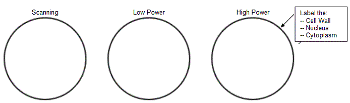
Part A - Onion Cells
Obtain a prepared slide of onion cells or prepare one yourself. View under the microscope and sketch the cells at each magnification. Label the cells as they appear under high power.
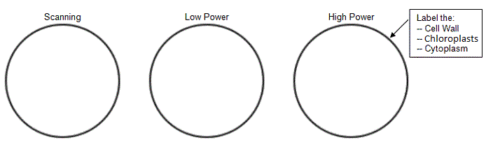
Part B - Elodea Cells
View a prepared slide of elodea (anacharis), which is an aquarium plant. As the slide warms from the light of the microscope, you may see the chloroplasts moving, a process called cytoplasmic streaming.
Post Lab Questions
1. Describe the shape and the location of chloroplasts.
2. Why were no chloroplasts found in the onion cells? (hint: think about where you find onions)
3. Which type of cell was smaller - the onion cells or the elodea cells? How do you know?
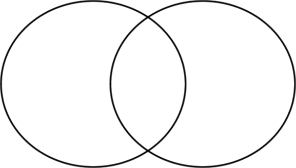
4. Fill out theVenn Diagram below
to show the differences and similarities between the onion cells and the elodea
cells.
5. Did you notice the chloroplasts moving within the cytoplasm of the elodea plant? Do they all move in the same pattern or direction?
Suggest a reason why these structures move.
Develop a quick experiment to test your hypothesis. Describe the test below, and if you have time conduct the test.