Rat Dissection - Organs of the Abdomen and Thoracic Cavities
1. Locate the diaphragm and the heart is centrally located in the thoracic cavity. The thymus gland, may be visible at the upper part of the heart.
The thymus gland is a small, bean-shaped organ located in the upper chest, in front of the heart. It is part of the lymphatic system, which helps the body fight infection. The thymus gland produces white blood cells called T cells, which help the body fight off infection. It also helps to regulate the immune system.
2. The lungs are spongy organs that lie on either side of the heart and should take up most of the thoracic cavity.
The lungs are two spongy organs in the chest that take in oxygen from the air and deliver it to the bloodstream. They also remove carbon dioxide from the bloodstream and release it into the air.
The lungs are made up of millions of tiny air sacs called alveoli. The alveoli are surrounded by a network of tiny blood vessels called capillaries. When you breathe in, oxygen from the air passes through the alveoli and into the capillaries. The oxygen then travels through the bloodstream to all parts of the body.
3. A sheet of muscle can be found just under the heart (and above the liver) - this is the diaphragm. This muscle is only found in mammals. (The diaphragm may have been cut when you opened the thoracic cavity.)
The Abdominal Organs
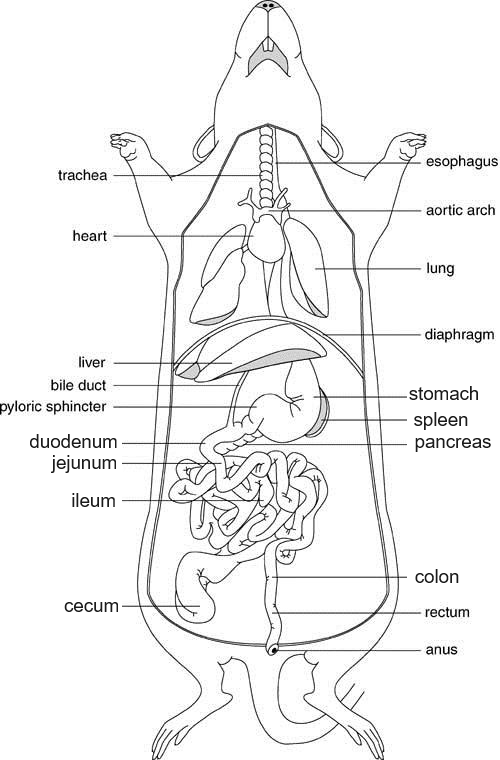
1. Locate the liver, which is a dark colored organ suspended just under the diaphragm. It has four lobes:
median or cystic lobe - located at the top, there is an obvious central cleft
left lateral lobe - large and partially covered by the stomach
right lateral lobe - partially divided into an anterior and posterior lobule, hidden from view by the median lobe
caudate lobe - small and folds around the esophagus and the stomach, seen most easily when stomach is raised
2. Find the stomach, its a curved organ lying just under the liver. At the top of the stomach you can see the esophagus where it pierces the diaphragm and joins the stomach. Lifting the stomach up may reveal a bumpy glandular organ: the pancreas.
3. The spleen is about the same color as the liver and is attached to the greater curvature of the stomach.
The spleen controls the level of white blood cells, red blood cells and platelets (small cells that form blood clots). It screens the blood and removes any old or damaged red blood cells. If the spleen doesn't work properly, it may start to remove healthy blood cells.
4. The small intestine is a slender coiled tube that receives partially digested food from the stomach (via the pyloric sphincter). It consists of three sections: duodenum, jejunum and ileum, (Listed in order from the stomach to the large intestine.)
The duodenum is the first part of the small intestine and is where bile and pancreatic juices are added to the food. These juices help to break down the food so that it can be absorbed by the body. The jejunum is the middle part of the small intestine and is where most of the nutrients from food are absorbed. The ileum is the last part of the small intestine and is where bile salts and vitamin B12 are absorbed.
5. Locate the colon, which is the large greenish tube that extends from the small intestine and leads to the anus. The colon is also known as the large intestine and it consists of four sections:
cecum - large flattened sac in the lower third of the abdominal cavity, it is a dead-end pouch and is similar to the appendix in humans.
ascending colon – food travels upward.
transverse colon – a short section that is parallel to the diaphragm
descending colon – the section of the large intestine that travels back down toward the rectum.
rectum - the short, terminal section of the colon that leads to the anus. The rectum temporarily stores feces before they are expelled from the body.
Rat Navigation
Step 1: Body Regions
Step 2: External Features
Step 3: Expose the Muscles
Step 4: Expose the Bones
Step 5: Head & Neck
Step 6: Thoracic & Abdomen
Step 7: Urogenital System