Cow Eye Virtual Dissection
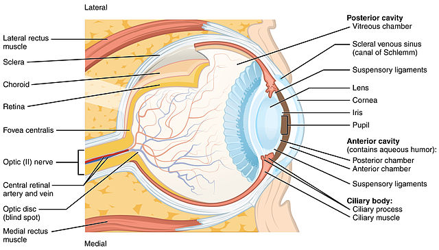
Diagram of the Eye
Procedure
1. Cut the fat around the eye.
2. Puncture the cornea with scissors or a scalpel. The cornea is pinned here.
3. Cut the eye into a front and a back half.
4. Open the eye. The gelatinous fluid inside is the vitreous humor, the lens sits within this liquid.
5. Separate the parts of the eye. The lens is the hard, sphere-shaped structure sitting on (or in) the vitreous humor.
6. On the back of the eye, the thin layer of cells of the retina can be seen here, it is very thin and easy to pull away. The spot where they remain attached is the optic disk, which connects the retina to the optic nerve.
7. The tapetum is clearly visible with the retina removed. It is blue and shiney and will reflect light. This helps a cow to see in the dark. Notice on this photo, you can see the optic disk, the spot where the retina is still attached.
8. This student has carefully removed the iris, which can be difficult to get out in one piece. The hole in the center of the iris is the pupil.
9. These eye parts are separated so that you can clearly see the main structures.
10. The optic nerve can be seen on the back side of the eye, the nerve is usually buried within the fat, so it takes some work to find it and expose it.