Cat Dissection Student Guide
Cat Dissection Student Handout (Google Doc)
Safety Protocols
- Wear safety goggles, the fluid used to preserve cats is toxic and can injure the eyes.
- Always clean your station and store cats in marked containers
- Wash hands, even if you did wear gloves
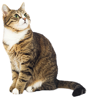
1. Remove your cat from the blue container marked with your hour.
2. Cut open one end of the bag and drain the excess fluid. – Save the bag, you will store your cats in it.
3. Take this time to label the bag with a permanent marker. Place at least ONE name from your group on the bag. This will ensure your cat will be reunited with your group should it become lost.
4. Cover a tray with paper towels and place the cat on the tray. This will make clean up easier
5. You are now ready to begin trying to locate and identifying the structures. Read instructions carefully, do not remove structures unless you are told to do so.
**Use the colored pins to mark structures as you find them** One member of your group will upload these photoes.
6. When there are about 7-8 minutes left in the period, you should begin cleaning up and putting your cats away. Put a rubber band around the end of the bag (we don’t want to “let the cat out of the bag”) and put the cats back in your hour’s blue container.
7. CLEAN everything. Wipe off dissecting trays and countertops, put equipment back where you got it.
8. If you miss any days, there are photos of the cat posted at biologycorner.com. Use these to catch up or to study. The lab will be open in the morning before school. Plan to be there is you miss a day.
9. Assessment for the cat dissection includes:
- Completion of the lab guide
- Attendance and make-up (in the morning)
- Uploaded photos
- Final Lab Test (either a practical or slides)
Muscles of the Cat
When you remove your cat from the bag, note that the skin has been removed and the superficial muscles are exposed. Your job will be to use the manuals provided to locate each of the muscles listed below. Check the box when you have located and pinned each of the structures.
Ventral Muscles
1. Sartorius
2. Gracilis
3. External Oblique
4. Latissimus Dorsi (can be seen partially)
5. Xiphihumeralis
6. Pectoralis minor
7. Pectoralis major
8. Pectoantebrachialis
9. Sternomastoid
10. Clavobrachialis
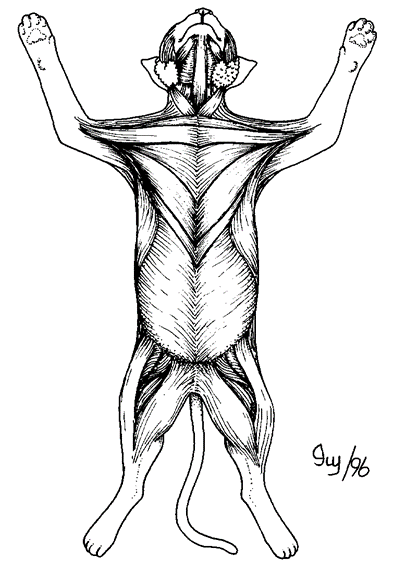
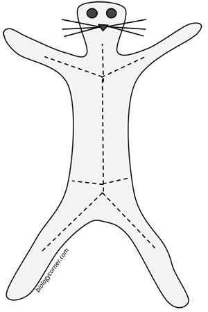
Label the muscles on the cat diagram:
Upload a photo of your cat with at least 5 ventral muscles labeled.
Dorsal Muscles
1. Clavotrapezius
2. Acromiotrapezius
3. Spinotrapezius
4. Latissimus Dorsi
5. External Oblique (partial)
6. Triceps Brachii (long head)
7. Triceps Brachii (lateral head)
8. Sartorius
9. Biceps Femoris
10. Gastrocnemius
11. Achilles tendon
Levator Scapula (a)
Spinodeltoid (b)
Brachioradialis (c)
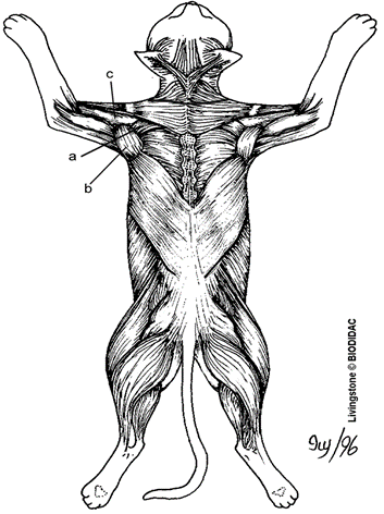
Label the muscles on the cat diagram:
Upload a photo of your cat with at least 5 ventral muscles labeled.
Blood Vessels above the Diaphragm
Double-injected cats are usually used to identify blood vessels. Arteries are injected with red latex, and veins are injected with blue latex. Blood vessels differ slightly in location from cat to cat. Carefully remove the fascia with blunt instruments to separate blood vessels from other structures.
 1. Place your cat in a dissecting tray with the ventral surface facing upward. Use a sclpel to make a Y incision in the thoracic cavity, then continue to the abdominal cavity as shown in the diagram. You may need to use pins to hold the skin open for viewing.
2. Identify the following major organs: heart, trachea, lungs, diaphragm, stomach, spleen, liver, small intestine, and large intestine. Use a lab manual to assist you in locating these structures. You do not need to pin them at this point and take care that you do not damage or remove them until you are instructed to (later in the lab.)
3. Using your scissors, cut open the pericardial sac surrounding the heart to expose the heart and the attached vessels. Identify the atria and ventricle of the heart.
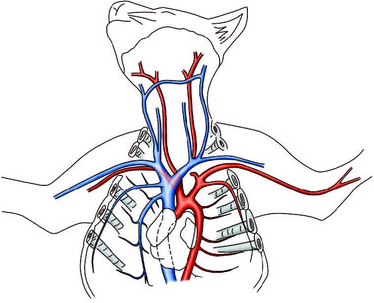
4. Refer to your lab manual to identify the following arteries that are located above the diaphragm. Use colored pins to mark the locations as you find them.
5. Identify the pulmonary trunk exiting from the right ventricle. Locate its branches, the right pulmonary artery and the left pulmonary artery, and follow them to the lungs. Pin this structure.
6. Identify the ascending aorta as it exits the left ventricle follow it to the aortic arch and trace its path into the abdominal cavity where it is referred to as the descending aorta and the abdominal aorta when it is below the diaphragm..
7. In cats, there are only two branches off the aortic arch, the brachiocephalic artery (first branch) and the left subclavian artery. Locate and pin those structures.
8. The brachiocephalic artery divides into the right subclavian artery, the right common carotid, and the left common carotid. Locate and pin these structures.
9. The superior vena cava can be seen on the top surface of the heart near the aorta.
10. Lift the heart aside to look behind it and find the inferior vena cava. Both of these veins return blood to the heart. Pin both the superior and inferior vena cava.
11. The superior vena cava splits into the left and right brachiocephalic veins. Pin both.
12. The jugular veins will join the brachiocephalic veins and drain blood from the head region. Pin the external jugular vein. (The internal may be difficult to locate.)
13. Color code the image below (red for arteries. blue for veins, and label each of the vessels in bold above.
14. Photograph your pinned cat with a key to the structures (labeled) and upload.
Vessels below the Diaphragm
Note: many of these vessels will be found by locating the organ they are attached to. Do not remove organs, instead, gently push them aside and tease away tissue that might be obscuring your view.
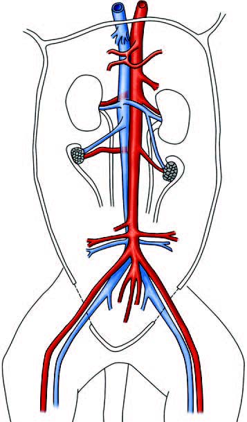
1. Lift the heart and follow the aorta until it goes through the diaphragm and becomes the abdominal aorta. All vessels you will locate will be directly attached to it, it is the largest artery in the body. 2. The inferior vena cava runs parallels to the abdominal aorta. Pin the inferior vena cava and the aorta. 3. The first branch (below the diaphragm) is the celiac trunk. This small artery then splits into three smaller branches: the hepatic artery which goes to the liver, the gastric artery that goes to the stomach, and the splenic artery that goes to the spleen. Pin the celiac artery and find its branches. 4. Just below the celiac trunk is the superior mesenteric artery which supplies blood to the mesentery of the small intestine. This artery is small and easily broken if you are too rough with the intestines. Pin it. 5. Tracing the aorta downward, you will find the renal arteries which are attached to the kidneys. The veins are near them and distinguished by a blue color. Pin the renal arteries/veins. 6. You may also be able to locate the gonadal arteries near the renal arteries. They supply the testes in males and the ovaries in females. 7. Farther down the aorta, you can find the inferior mesenteric artery. It is also small and fragile and may broken due to moving the intestines around. Place a pin in it. 8. Continue to trace the aorta toward the legs. Eventually it will split and form a Y, with the left external iliac going to the left leg and the right external iliac going toward the right leg. The internal iliac artery will go straight toward the tail. Pin each of these arteries. 9. In this area, you will also see the external iliac veins, it will be blue and run parallel to the external iliac artery. Pin one. 10. Trace the external iliac artery into the leg where it will become the femoral artery. Next to it will be the femoral vein. Pin both. 11. Color code the diagram below (the aorta is the large vessel on the right) with red for artery and blue for vein. Label each of the bold structures you found above.
12. Have your instructor check the pins for the vessels associated with the heart. Instructor initials ___________
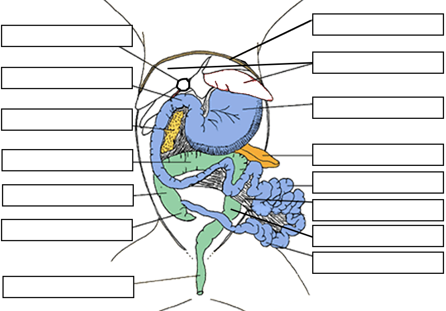
Gross Anatomy of the Cat
In this investigation you will explore the abdominal and thoracic cavity of the cat. A careful dissection will reveal structures we have learned in class. Follow the directions step-by-step and answer or sketch where asked. Return any organs you remove to the cat, you made need them for the final lab test.
1. A curtain of fat lies over most of the organs of the abdominal cavity, this is called the greater omentum. It can be removed so that organs are visible. Describe this structure.
2. The liver is the largest organ in the abdominal cavity, and it is divided into lobes. How many lobes of the liver do you count? Sketch and label them.
3. Push the liver upward to locate the gallbladder that lies underneath and find the bile duct, which connects the gallbladder to the duodenum of the small intestine. Sketch each of these structures.
4. Locate the diaphragm which lies above the liver and separates the abdominal cavity from the thoracic cavity. Describe the appearance of the diaphragm, to what body system does it belong?
5. The lungs are located on either side of the heart in the thoracic cavity. Are the lungs superior (above) or inferior (below) to the diaphragm?
6. The lungs connect to the trachea, or the airway. It has cartilage rings that give it a ridged appearance and keep it from collapsing. Locate the trachea and follow it toward the mouth. You will eventually find a widened muscular area, the larynx, which is the cat’s voice box (or “meow box”). What do you think will happen to the lungs if air is blown into the trachea? (Your instructor may demonstrate this for you.)
7. Locate the point at which the esophagus pierces the diaphragm (the hiatus) and trace the esophagus to the stomach. Wiggling the stomach may help you find the esophagus. Trace it upwards to where it is visible near the trachea. Describe the difference between the esophagus and the trachea. How can you tell which is which?
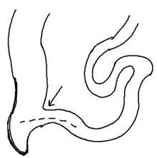
8. Remove the stomach and inspect the contents. Often these cats are carrying parasites and can be seen within the stomach or intestines. Describe (or sketch) the inside of the stomach, paying attention to its texture. Investigate the shape of the cardiac sphincter valve and the pyloric sphincter valve. Sketch the stomach, labeling the lesser and greater curvature, the esophagus, and the two sphincter valves.
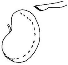
9. Push the stomach aside to locate a bumpy structure underneath it, the pancreas. You can also find the spleen laying over the top of the stomach in this area. Describe the appearance of the spleen
10. Locate the duodenum of the small intestine, it will be a straight section of just after the stomach. Locate the jejunum and the ileum and note where the ileum joins the large intestine at the ileocecal valve.
11. You should also be able to find the cecum in this area as a pouch attached to the large intestine. Label each of these structures of the drawing.
12. Stretch the jejunum out so that the membrane holding it together is visible. The mesentery should have small vessels visible within it, these mesenteric vessels connect to the superior and inferior mesenteric arteries.
Compare the mesentery to the omentum. Imagine you are describing these two membranes to a person not familiar with the cat; how would you explain how they are different?
13. Examine the large intestine closely. In cats, the ascending, transverse, and descending colon are present, but much shorter than what is seen in humans. Avoid cutting the rectum of the cat, this will likely contain feces.
Sketch and label these structures:
14. At this point, you can remove the small intestine, cutting it at the duodenum and the ileocecal valve. Cut the mesentery so that it can be measured.
What is the length of the small intestine? _______ cm
What is the length of the large intestine (does not need to be removed) _______ cm
15. Trace the ureters from each kidney. Wiggling the kidneys make help you locate this tube.
Do the ureters enter the bladder at the same spot? _______
Remove the bladder and fill it with water. Is it waterproof? _________
16. Describe the location of the kidneys. Kidneys are said to sit “retroperitoneally”. What does this mean?
Remove one of the kidneys. What vessels must you cut in order to remove the kidney?
Make a longitudinal cut in the kidney and view the cortex and medulla. Draw the renal pyramids. How many are there? ________
17. If your cat is a female, locate the uterine horns, the ovaries and the vagina.
If your cat is a male, locate the testes (if present) and the penis. Find a cat of the opposite sex to see structures your cat doesn't have.
Where does the urethra exit the body in the male compared to that of a female cat? Is it possible to tell the difference between males and females from an external view? How?
18. Label the image.
Structure Checklist - Can you briefly describe the location of each of the following structures?
Respiratory System Trachea Left and Right Lung Larynx Diaphragm
Circulatory System Pulmonary Artery Aorta Heart (atrium/ventricle) Vena Cava
Reproductive / Urinary System Testes Ovary Uterine Horn Vagina Urinary Bladder Ureter Kidney
Digestive System
Greater Omentum Mesentery Stomach Liver Esophagus Gallbladder Pancreas
Spleen Bile Duct Cecum Colon Rectum Small intestine (duodenum, jejunum, ileum)


 This work is licensed under a Creative Commons Attribution-NonCommercial-ShareAlike 4.0 International License.