REVIEW GUIDE - CARDIOVASCULAR SYSTEM
1. How do you calculate cardiac output (formula)? heart rate x stroke volume
2. Arteries carry blood [ away from / toward ] the heart.
Veins carry blood [ away from / toward ] the heart.
[ Arteries / Veins ] carry oxygenated blood? _
Name the one vessel that is the exception: pulmonary artery
3. What do you call the loose, outer layer of the sac around the heart?
pericardium
What is the inner layer? visceral Outer? _parietal _
4. When vessels expand, it is called __vasodilation____
When they contract? __vasoconstriction ________
5. Describe the epicardium, myocardium, and endocardium
epicardium = outer layer
endocardium = inner layer, lining the chambers of the heart
myocardium = muscle layer of the heart
6. Describe the size and location of the heart. size of a fist, center of chest, within the mediastinum
What is the apex of the heart and where is it located? point of the heart
What divides the left and right side of the heart? ___septum____
Which side is more muscular? ___ left side________ What bone lies directly in front of the heart? __sternum____
7. During systole, the aortic valve is _____open_____.
During diastole, the aortic valve is _____closed_____.
8. Veins and arteries meet at ___capillaries__, where nutrients are exchanged with body tissues.
Arteries branch into smaller vessels called _____arterioles_
Veins also have smaller branches called venuoles
9. What aids in bringing blood back to the heart? diaphragm, sphincter valves, movements of skeletal muscles
10. Trace the path of a nerve (cardiac) impulse through the cardiac conduction system.
What is the pacemaker? ___SA node, regulates the pace of the heart ___
What fibers cause a contraction in the ventricle? Perkinje Fibers
Where is the AV node located? between right atrium and ventricle of the heart (AV stands for atrioventricular)
11. What do the terms tachycardia and bradycardia mean? What is arrhythmia? What is defibrillation?
tachycardia = rapid heart rate; bradycardia = slow heart rate; arrhymia = irregular heart rate
a defibrillator shocks the heart back to its normal rhythm
12. How are the valves attached to the wall of the heart (2 structures)? chordae tendinae attached to papillary muscles
13. Generally speaking, when the ventricle contracts, the atrium relaxes
Any contraction (atrial or ventricular) is called __systole_ Relaxing is called _____diastole __.
14. What causes a P-Wave, the QRS complex and a T wave?
P is the first small bump, QRS is the large peak, and T is the small wave after the peak
What is an ECG? electrocardiogram * Be able to analyze one on the test.
15. What are systolic pressure and diastolic pressure? What is the “normal” blood pressure for a human?
16. What two pieces of equipment are needed to take a person’s blood pressure?
Describe the procedure: the stethoscope is places at the brachial artery (elbow) and the cuff is wrapped around the arm, the cuff is inflated and then the valve is released slowly. The first time you hear the sound of a heart beat is the systolic pressure. The cuff continues to deflate until you no longer hear the sound, this is the diastolic pressure.
17. Blood that moves from the heart to the lungs and back to the heart again is in the ___pulmonary___ circuit.
Blood moving throughout the body is in the _systemic_ circuit.
18. Identify the three major vessels that branch off of the aortic arch. You may want to draw a diagram. brachiocephalic, left common carotid, left subclavian
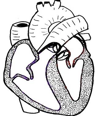
19. Label the heart and trace the flow of blood through the major vessels and all of the valves. (See image above)
20. Know the major disorders of the heart that we discussed in class and describe the treatment for each.
 This work is licensed under a Creative Commons Attribution-NonCommercial-ShareAlike 4.0 International License.