Chapter 13 – Cardiovascular System
13.1 Introduction
Systemic Circulation – delivers blood to all body cells and carries away waste
Pulmonary Circulation – eliminates carbon dioxide and oxygenates blood (lung pathway)
Arteries - carry blood away from the heart
Veins - return blood to the heart
13.2 Structure of the Heart
Heart Size – about 14 cm x 9 cm (the size of a fist)
Coverings of the heart
Pericardium encloses the heart (like a bag) (visceral, parietal)
Pericardial cavity – contains fluid for the heart to float in, reducing friction
Wall of the Heart
Epicardium – outer layer, reduces friction
Myocardium – middle layer, mostly cardiac muscle
Endocardium – inner layer, blood vessels and Purkinje fibers
13.3 Heart Chambers and Valves
Atria - top chambers
Ventricles - bottom chambers
Septum - divides left and right side
Atrioventricular Valve (AV) - these valves are located between the atrium and the ventricle
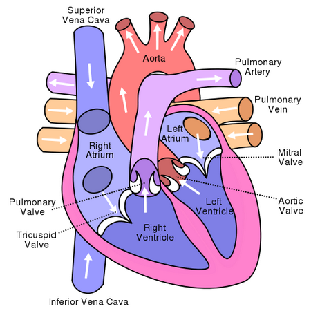
Tricuspid - right side AV
Bicuspid - left side AV, also called mitral valve
Superior Vena Cava - vessel the returns blood to the heart from the upper body
Inferior Vena Cava - vessel the returns blood to the heart from the lower body
Coronary Sinus - indentation on the front of the heart
Chordae tendinae / Papillary Muscles - muscles and tendons that hold the heart valves in place
Pulmonary Trunk/Arteries - large vessel that splits into the left and right pulmonary arteries, these are the only arteries that carry deoxygenated blood
Pulmonary valve - controls the flow of blood into the pulmonary trunk
Pulmonary Veins - returns oxygenated blood from the lungs
Aorta - large vessel that delivers blood to the body
Aortic Valve - controls the flow of blood into the aorta
Path of Blood Through the Heart
Quick Overview
1. Deoxygenated blood enters right atrium through the vena cava
2. Blood moves into the right ventricle
3. Blood goes out the pulmonary arteries and heads to the lungs
4. Blood returns from the lungs and enters the left atrium
5. Blood moves into the left ventricle
6. Oxygenated blood moves out of the left ventricle through the aorta and to the body
Labeling Practice - External Heart
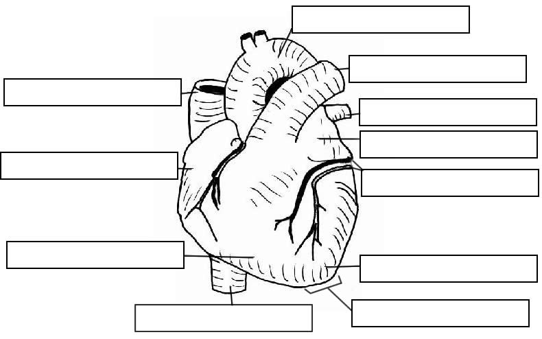
See Also: Labeling Internal Heart

.svg)