Cat Dissection Questions
1. How many lobes of the liver do you count? Use your book or other resources to sketch the liver and name each of the lobes.
What organ is found embedded within the liver?
Carefully remove the entire liver. Turn it over to view the bile duct that connects the liver to the duodenum.
2. How many lobes of the lung do you count on the left side? ____ On the right side? ______
How does this compare to the number of lobes on a human?
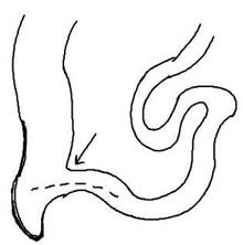
3. With the liver removed, you should be able to see the stomach. Identify the greater and lesser curvature. The stomach connects to the esophagus at its upper end. Once you have located the esophagus, cut a hole in it and insert a blunt probe, slowly push the probe into the esophagus until you reach the stomach.
You can use the probe to discover the location of the esophageal hiatus. The place on the diaphragm where the esophagus pierces it.
Use a ruler to measure the length of the esophagus on the cat. ___________
4. Compare the esophagus to the trachea. Clear the tissue around the trachea to locate the larynx.
Describe the difference between the esophagus and the trachea. How can you tell which is which?
5. The lower end of the stomach connects to the duodenum of the small intestine. This is a short, straight section just after the stomach. The second section is called the jejunum, which is held together by a membrane called the mesentery.
Compare the mesentery to the omentum. Imagine you are describing these two membranes to a person not familiar with the cat; how would you explain how they are different.
6. Continue to trace the small intestine until it joins with the large intestine. The last section of the small intestine is called the ileum.
How can you tell the difference between the large and small intestine on the cat?
Sketch the 3 sections of the small intestine and indecate where you find the mesentery.
 7. Locate the cecum. The valve between the cecum and the ileum is the ileocecal valve. Label the image
8. Trace the large intestine as it goes up and then makes a U-turn to leave the cat at the rectum. The three sections are the acending colon, transverse colon, and descending colon.
Sketch the large intestine and label each section.
9. Locate the spleen and the pancreas. Describe how their appearance differs.
10. Find the diaphragm. Compare the appearance of this muscle to a skeletal muscle, like the kind found in the leg.
11. Trace the ureters from each kidney. Wiggling the kidneys may help you locate these small tubes.
Do the ureters enter the bladder at the same spot? _______
Remove the bladder and fill it with water. Is it waterproof? ________
 12. Describe the location of the kidneys. Kidneys are said to sit “retroperitoneally”. What does this mean?
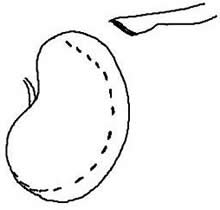
Remove one of the kidneys. What three structures must you cut in order to remove the kidney?
Make a longitudinal cut in the kidney. Sketch below and label the renal pyramids, cortex, and medulla.
13. Remove the small intestine and cut the mesentery so that the intestine can be stretched out.
With a ruler, measure the length of the small intestine: _______
Measure the length of the large intestine (you do not need to remove it.) __________
14. Remove the stomach and inspect the contents. Often these cats are carrying parasites that can be seen within the stomach or intestines. Describe (or sketch) the inside of the stomach, paying attention to the texture of the rugae. Investigate the shape of the cardiac sphincter valve and the pyloric sphincter valve.
Sketch the stomach, label the lesser and greater curvature, the esophagus, and the two sphincter valves.
15. Carefully remove the heart and the lungs. Lay the heart and lungs on a paper towel and find the: superior, inferior, and middle lobes. Sketch and label below.
16. Cut the heart in half and observe the chambers of the heart. Sketch and label the ventricle and atria.
17. If your cat is female, locate the uterine horns, the ovaries, and the vagina. Sketch and label below. (If you do not have a female, locate one of the room to view.)
If your cat is a male, locate the testes (if present) and the penis. You do not need to sketch this. (If you do not have a female, locate one to view.)
Where does the urethra exit the body in the male compared to the female cat. Is it possible to tell the difference between males and females from an external view? How?
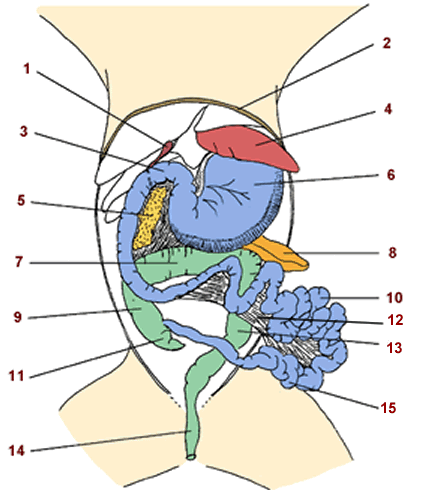
18. Label the structures:
Checklist - you should be able to identify each of the following structures on a photo, diagram, or cat specimen.
Respiratory System
![]() Trachea
Trachea ![]() Left and Right Lung
Left and Right Lung ![]() Larynx
Larynx
![]() Heart
Heart ![]() Diaphragm
Diaphragm ![]() Pulmonary Artery
Pulmonary Artery ![]() Aorta
Aorta
Reproductive / Urinary System
![]() Testes
Testes ![]() Ovary
Ovary ![]() Fallopian Tube / Uterine Horn
Fallopian Tube / Uterine Horn ![]() Vagina
Vagina
![]() Urinary Bladder
Urinary Bladder ![]() Ureter
Ureter ![]() Kidney
Kidney ![]() Ureter
Ureter
Digestive System
![]() Greater Omentum
Greater Omentum ![]() Mesentery
Mesentery
![]() Greater and Lesser Curvature (stomach)
Greater and Lesser Curvature (stomach) ![]() Rugae
Rugae
![]() Fundus of Stomach
Fundus of Stomach ![]() Esophagus
Esophagus ![]() Hiatus
Hiatus
![]() Liver (right medial, right lateral, left medial, left lateral)
Liver (right medial, right lateral, left medial, left lateral)
![]() Gall Bladder and Bile Duct
Gall Bladder and Bile Duct ![]() Pancreas
Pancreas ![]() Spleen
Spleen ![]() Cecum
Cecum
![]() Colon (ascending, transverse, descending)
Colon (ascending, transverse, descending) ![]() Rectum / Anus
Rectum / Anus
![]() Small Intestine ( duodenum, ileum, jejunum)
Small Intestine ( duodenum, ileum, jejunum)
 This work is licensed under a Creative Commons Attribution-NonCommercial-ShareAlike 4.0 International License.