Cat Dissection Image Gallery
Cat Images are Located in this folder.
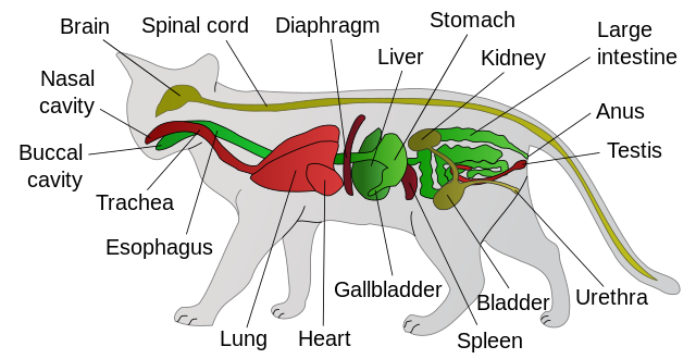
Classroom Resource: Pictorial Anatomy of the Cat
1. The largest organ is the liver, and seen embedded within the right medial lobe is the gall bladder. Not seen on this photo is the right lateral lobe which lies to the side of (and behind) the right medial lobe. The caudate lobe can also be seen by lifting the left lateral lobe.
2. When you lift the liver, the stomach is revealed as a pouch underneath. The first part of the small intestine in the duodenum, shown on the photo attached to the stomach. The second section of the small intestine in the jejunum.
3. From a different angle, you can see how the stomach connects to the esophagus. In this image, a blunt probe was used to trace the esophagus all the way to where it pierced the diaphragm at the hiatus and entered the stomach.
4. If you trace the small intestine to the large intestine, you will find the cecum, which is a pouch-like structure at the beginning of the ascending colon. The valve between the large and small intestine is called the ileocecal valve.
5. There are two membranes that are visible in the abdominal cavity of the cat. The greater omentum is a fatty membrane that lies on top of the organs of the digestive system (1st photo). The mesentery holds the coils of the small intestine together and has easily visible mesenteric arteries (2nd photo).
6. The spleen is visible as a dark brown structure that lays over the top of the stomach an intestines.
7. The pancreas lies under the stomach and the spleen, you will probably need to life the stomach to find it. One of the distinguishing features of the pancreas is its bumpy texture.
8. To identify the sections of large intestine, it may be necessary to remove the small intestine. Shown below is the cecum, ascending colon, transverse colon, and descending colon (rectum).
9. The kidney is located behind the peritoneum (retroperitoneally) and shown below with the membrane removed. The ureter is a pale tube attached to the kidney and the renal vein is visible as a blue structure.
 This work is licensed under a Creative Commons Attribution-NonCommercial-ShareAlike 4.0 International License.