Virtual Lab - Amoeba and Paramecium
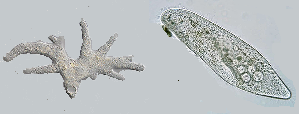
The amoeba is a single-celled organism with a flexible cell membrane and no fixed shape. It moves and feeds through the formation of pseudopodia, which are temporary projections of its cytoplasm. Amoebas move by extending their pseudopodia and flowing into the direction of their movement.
The paramecium is also single-celled organisms but have a more defined shape, resembling a slipper. They possess cilia, which are hair-like structures used for movement and feeding. Paramecia move using their cilia, which beat in a coordinated manner, allowing them to move more rapidly and in a directed manner compared to amoebas.
Both protists are found in fresh water, like ponds and streams. They are heterotrophs, meaning they must consume food. The amoeba feeds by engulfing food with their pseudopodia. Paramecia sweep food paricles into their oral groove.
Ameboid Movement -
Access the media at: biol.co/virtameb - this will take you to a flickr album.
1. View the slides that show an ameba consuming a small ciliate.
2. Go to youtube and watch the video.
Paramecium
View the video at youtube showing how paramecium move.
Analysis
1. Describe how the paramecium eats and digests food:
2. Describe how the amoeba eats and digests food.
3. Compare the movement of the paramecium to the movement of the ameba.
4. How are the paramecium and the ameba alike?
5. How are the paramecium and the ameba different?
6. Compare the shape of the paramecium to the shape of the ameba.