Review Guide - TISSUES
Review the Slides for this unit.
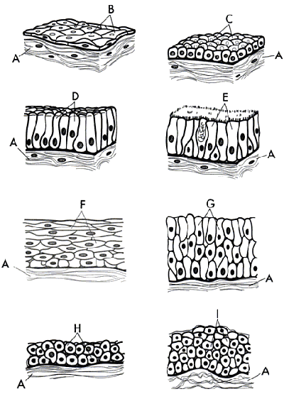
1. Identify the types of tissues shown below.
What do each of the letters represent?
2. List the four types of tissues and describe their general function / location.
3. Identify the types of epithelial tissues from pictures using the naming conventions (simple, stratified, cuboidal, squamous, columnar)
4. Know the general locations of epithelial tissue (stratified squamous, transitional, simple squamous, stratified columnar)
5. Compare the appearance and function of cilia and microvilli.
6. Define and locate the basement membrane on a picture.
7. Describe the matrix. (See Connective Tissue Coloring). Know what each part of the matrix does and what it looks like. - macrophages, mast cells, fibroblasts, elastic fibers, collagen fibers, ground substance). Be able to label a picture of the matrix.
8. Compare loose connective tissue to fibrous (dense) connective tissue. Where is each found?
Distinguish between a tendon and a ligament
9. Describe adipose tissue.
10. Compare the main types of cartilage hyaline, elastic, and fibrocartilage. Know where each is located within the body.
11. List the three types of muscles tissue and know where they are found. Which ones are voluntary and which ones are involuntary?
12. What is the main cell of the nervous tissue? What is an support cell found within nervous tissue?
13. Describe the properties of skin, focusing on skin color and the cells that contain pigments (melanocytes). Discuss how skin color provides evidence of evolution in humans. See HHMI: The Biology of Skin Color
14. What is epidermolysis bullosa - be able to describe the disease, its causes, and its relationship to the epidermal and connective tissues.