Student Notes: Sense of Sight
Sensory Perception - Vision
Student slides and notes covers aspects of vision and visual processing.
Anchoring phenomenon is a case about a congenital case of blindness called leber amaurosis.
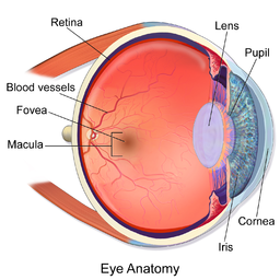
Eye Anatomy
-
Cornea: The transparent, dome-shaped outer surface that covers the front of the eye. It helps in focusing light.
-
Pupil: The black circular opening in the center of the iris that allows light to enter the eye.
-
Iris: The colored part of the eye that surrounds the pupil. It controls the size of the pupil and hence regulates the amount of light entering the eye.
-
Lens: Behind the iris, the lens focuses light onto the retina. It adjusts its shape to allow the eye to focus on objects at different distances.
-
Retina: The innermost layer at the back of the eye. It contains photoreceptor cells—rods and cones—that convert light into electrical signals and send them to the brain through the optic nerve.
-
Optic Nerve: A bundle of nerve fibers that carries visual information from the retina to the brain, where it is interpreted.
-
Vitreous Humor: A gel-like substance that fills the space between the lens and the retina, helping the eye maintain its round shape.
-
Sclera: The white, tough outer layer of the eye that protects the eyeball.
-
Choroid: A layer between the retina and the sclera that contains blood vessels, providing nourishment to the eye.
-
Ciliary Body: Produces the aqueous humor (fluid) that nourishes the cornea and lens and helps maintain the shape of the eye.
- Aqueous Humor: A clear fluid that fills the space between the lens and the cornea, providing nutrients to these parts of the eye and maintaining their shape.