Study Guide - Musclular System
Muscular System Functions
- Movement: Facilitates body movement by contracting and relaxing muscles.
- Posture and Stability: Muscles provide support and maintain posture.
- Heat Generation: During muscle contraction, heat is produced, which helps regulate body temperature.
Skeletal Muscles
- Structure: Skeletal muscles are attached to bones by tendons. They're voluntary muscles, meaning they're under conscious control.
- Function: Responsible for movement by contracting and relaxing.
- Examples: Biceps, triceps, quadriceps, hamstrings.
Smooth Muscles
- Structure: Found in the walls of internal organs, blood vessels, and the digestive system. Involuntary muscles controlled by the autonomic nervous system.
- Function: Regulate involuntary movements like peristalsis (digestive movement) and control blood pressure.
- Examples: Muscles in the stomach, intestines, blood vessels.
Cardiac Muscles
- Structure: Unique to the heart, forming its walls.
- Function: Involuntary contraction to pump blood throughout the body.
- Characteristics: Striated like skeletal muscles but function involuntarily like smooth muscles.
Muscle Fibers:
Muscle fibers, also called muscle cells or myofibrils, are the individual cells that make up skeletal muscles. Myofibrils are bundled into groups called fascicles.
- Sarcolemma: The cell membrane of a muscle fiber.
- Sarcoplasm: The cytoplasm of a muscle fiber that contains organelles like mitochondria and the sarcoplasmic reticulum.
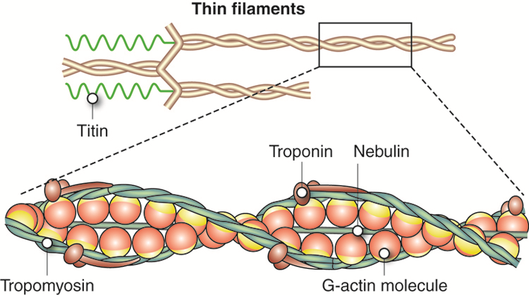
- Myofibrils: Thread-like structures within muscle fibers containing contractile units called sarcomeres. Consist of two types: thin (actin) and thick (myosin)
- Sarcomeres: Functional units of muscle contraction composed of actin and myosin filaments. This is the contracting unit of the muscle
Motor Unit
A motor unit consists of a motor neuron and all the muscle fibers it innervates (controls).The neuromuscular junction is crucial for the transmission of signals from the nervous system to muscle fibers, initiating the process of muscle contraction.
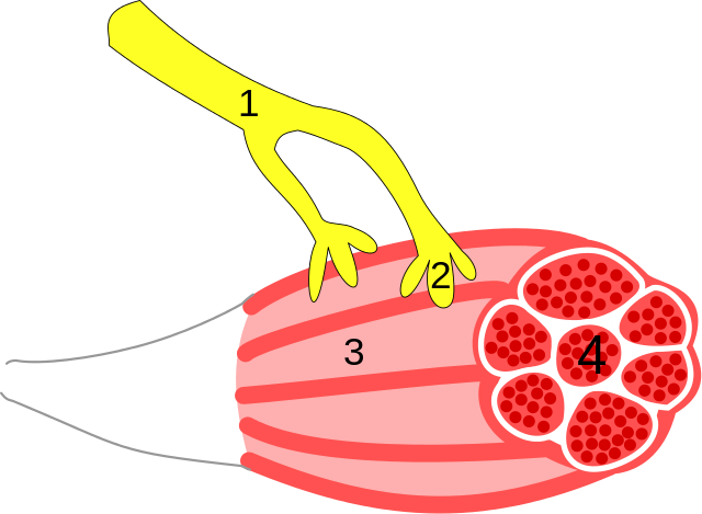
1) Axon 2) Motor End Plate 3) Muscle fiber 4) Myofibrils
- A motor unit consists of a motor neuron and all the muscle fibers it innervates (controls).
- Motor End Plate: Specialized region of the muscle cell membrane (sarcolemma) where the neuromuscular junction is located.
- Synaptic Cleft: Gap between the motor neuron and the motor end plate.
- Neurotransmitter: Acetylcholine (ACh) is released from the motor neuron and diffuses across the synaptic cleft to bind to receptors on the motor end plate.
Sliding Filament Theory
Describes the process of muscle contraction at the molecular level. Actin and myosin are the primary proteins involved in muscle contraction.
Key Steps in Muscle Contraction:
-
Resting State: When a muscle is at rest, actin and myosin filaments partially overlap but do not interact.
-
Initiation of Contraction: Upon receiving a signal from a motor neuron, calcium ions (Ca2+) are released from the sarcoplasmic reticulum (a specialized organelle in muscle cells).
-
Actin-Myosin Interaction: Calcium ions bind to troponin, causing a conformational change that moves tropomyosin away from the binding sites on actin.
-
Cross-Bridge Formation: Myosin heads (extensions of the myosin protein) attach to exposed binding sites on actin, forming cross-bridges.
-
Power Stroke: ATP (adenosine triphosphate) is hydrolyzed into ADP (adenosine diphosphate) and inorganic phosphate, releasing energy that causes the myosin heads to pivot, pulling the actin filaments towards the center of the sarcomere (the functional unit of muscle contraction).
-
Muscle Contraction: As long as calcium ions and ATP are present, the cycle of cross-bridge formation, power stroke, and release continues, causing the sarcomere to shorten, leading to muscle contraction.
-
Relaxation: When the nervous signal ceases, calcium ions are actively transported back into the sarcoplasmic reticulum, troponin-tropomyosin complex blocks the actin binding sites again, and the muscle relaxes.