Digestive System - Student Notes


Introduction
The digestive system serves the purpose of breaking down food into nutrients that the body can absorb, use for energy, and for building and repairing tissues.
Function: the mechanical and chemical breakdown of foods and the absorption of nutrients by cells
Consists of: alimentary canal ( 9 m from mouth to anus) and accessory organs
General Characteristics of the Alimentary Canal
(mouth, pharynx, esophagus, stomach, sm intestine, large intestine, anus)
Structure of the Wall of the Alimentary Canal
1. Mucosa (mucous membrane) – protects tissues, carries out absorption
2. Submucosa - contains glands, blood vessels, lymphatic vessels, nerves
3. Muscular layer – smooth muscle tissue, circular and longitudinal fibers, pushes food
4. Serosa (serous layer) – visceral peritoneum, outer covering of the tube, moistens and lubricates structures
Peristalsis- muscular contractions that move material through the alimentary canal
Mouth
Mouth – begins digestion by reducing size of particles (chewing) and mixing with saliva
Tongue – moves food during chewing, connects to the floor of the mouth via the frenulum, contains papillae (taste buds)
Palate – forms roof of oral cavity (hard and soft), uvula at back of the mouth
Palatine tonsils – back of the mouth/throat, organs that protect against infection
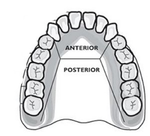
Teeth – primary vs. secondary teeth;
Anatomy of a Tooth
Crown – projects above the gums
Root – anchored to the alveolar process of the jaw
Enamel – made of calcium salts, hardest substance in body (outer surface)
Dentin – similar to bone, surrounds tooth’s central cavity
Blood vessels and nerves extend through the tooth via the root canal
Salivary Glands
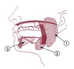
Serous cells produce amylase – splits starch and glycogen into disaccharides
Mucous cells produce mucus – lubrication during swallowing
1. Parotid Glands
2. Submandibular Glands
3. Sublingual Glands
Pharynx and Esophagus
Pharynx – nasal and oral cavitys - nasopharynx, oropharynx, laryngopharynx
Esophagus – moves to the stomach, penetrates the diaphragm at the esophageal hiatus
lower esophageal sphincter (cardiac sphincter) - prevent food and chemicals from moving up out of stomach
Stomach
*J-shaped, puchlike organs that hangs inferior to the diaphragm, 1 liter capacity
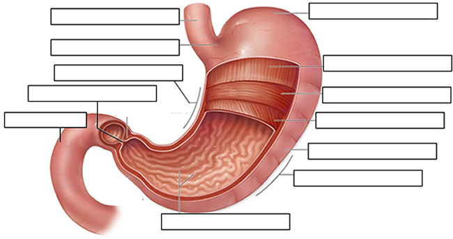
3 sets of stomach muscles: longitudinal, oblique, circular | Greater and Lesser Curvature
Four main parts of the stomach
1. Cardiac (esophageal opening, cardiac sphincter)
2. Fundic (temporary storage area, lies slightly above the cardiac region)
3. Body (central area of the stomach)
5. Pyloric (pyloric sphincter, controls emptying of the stomach into the sm. Intestine)
Lining of the stomach is a mucous membrane – with small openings called gastric pits, containing gastric glands
Gastric Juice - pepsin / intrinsic factor
Chyme – paste of food molecules after its been broken down by the movement of stomach and gastric juices, it is released from the pyloric sphincter valve into the first portion of the small intestine – duodenum
Rugae – folds within the stomach, increase surface area
Pancreas and Liver
has endocrine and exocrine functions - secretes pancreatic juice
Pancreatic juice – digests fats, breaks down nucleic acids into nucleotides
Liver - has large right and small left lobe
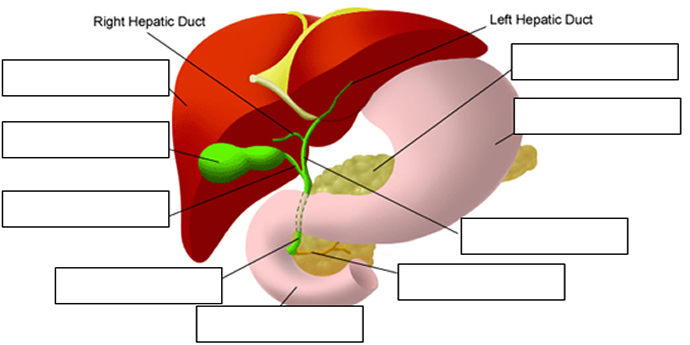
BILIARY SYSTEM – functions to create bile used in digestion; liver, gall bladder and ducts
Hepatic portal vein – delivers blood to the liver
Functions: maintains blood glucose, breakdown of lipids and fats, protein metabolism, stores iron and vitamins, destroys damaged red blood cells, removes toxic substances, secretes bile
Bile – yellowish-green liquid secreted from hepatic cells (when bile pigments build up in blood, skin turns green, a condition called jaundice). The hepatic duct joins the cystic duct to form the common bile duct, which empties into the duodenum
Bile aids in digestion, bile salts break down fat globules into smaller droplets – emulsification
Small Intestine
*tubular organ that extends from the pyloric sphincter, many loops and coils, fills much of the abdominal cavity
*receives secretions from the pancreas and liver, completes digestion of nutrients and chime, absorbs
1. Duodenum - first part of the small intestine
2. Jejunum – second part, ~2.2 m
3. Ileum – third part, longest ~3.3 m *jejunum and ileum are continuous
Mesentery – supports the coils of the small intestine, contains blood vessels to carry nutrients away
Greater Omentum – peritoneum membrane that drapes like an apron over parts of the system
Intestinal Villi – increase surface area for absorption
*the main function of the small intestine is to secrete chemicals to break down food and carry nutrients to blood (absorption)
Large Intestine
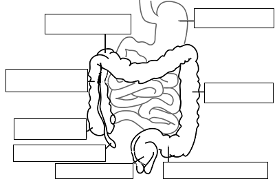
(named because its diameter is greater than the small intestine)
1. Cecum – beginning of the large intestine, pouchlike, closed end called the appendix (ileocecal valve)
2. Colon – ascending / transverse / descending / sigmoid
3. Rectum – stores waste before it is expelled from the body
4. Anus -muscular sphincter which controls the exit of waste
Functions –reabsorbs water and passes along material that was not digested; contains intestinal flora (bacteria to break down cellulose, also produce intestinal gas)
Mass movements – large portions of the colon contract to move material through it, usually after eating (defecation)
Disorders of the Digestive System
1. GERD (gastroesophageal reflux disease) / Heartburn
2. Diarrhea or Dysentery
3. Hepatitis (A, B, C)
4. Crohn’s Disease
5. Irritable Bowel Syndrome
6. Stomach Ulcers
7. Lactose Intolerance
8. Appendicitis
9. Hernia
10. Gallstones
11. Celiac Disease
12. Scurvy